Figures & data
Figure 1. (a) MRI (T1 FS SPIR sequence with contrast) of popliteal fossa before treatment. (b) Subtraction sequence with contrast directly post-HIFU. (c) Post-processing of lesion with tumor tracking tool. Note that due to (unfortunate) different angulation of MR-sequences pre- and post-contrast the bone is not in the same plane as the tumor in the pre-contrast sequence.
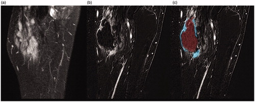
Table 1. Summary of patient data incl. prior treatments, effect of HIFU, and adverse effects.
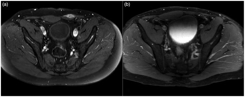
Figure 2. MRI (T1 FS SPIR sequence with contrast) of lower abdomen with desmoid within the left lower rectus abdominis muscle: (a) before treatment and (b) five years after treatment with no residual tumor.