
Figures & data
Figure 1. Transcervical injection of a dilute vasopressin solution into the space between the myometrium and surface of the leiomyoma through the leiomyoma using a PTC needle. My: myometrium; P: PTC needle; UC: uterine cavity; CL: cellular leiomyoma; Fa: feeding artery; Vp: vasopressin solution in the space between the myometrium and leiomyoma; Ab: ablation spot.
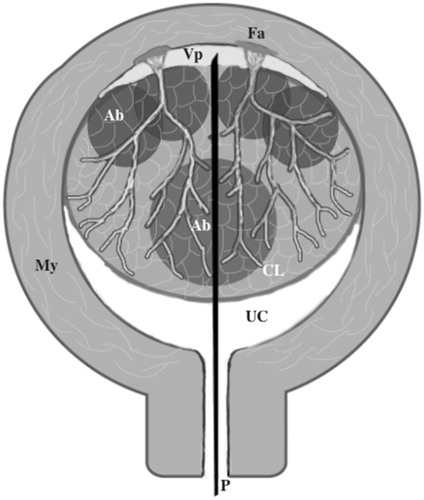
Figure 2. (a) T2-weighted magnetic resonance imaging (MRI) of a cellular leiomyoma (triangle) before transcervical microwave myolysis (TCMM). (b) Gadolinium-enhanced T1-weighted MRI of a cellular leiomyoma one month after TCMM & microwave endometrial ablation. The necrotic changes of the cellular leiomyoma are depicted as a de novo avascular area (triangle). The leiomyoma had shrunk from 5.6 cm to 3.4 cm in size. (c) Histological findings of a cellular leiomyoma on needle biopsy before TCMM. Stained with hematoxylin and eosin. Magnification ×200.
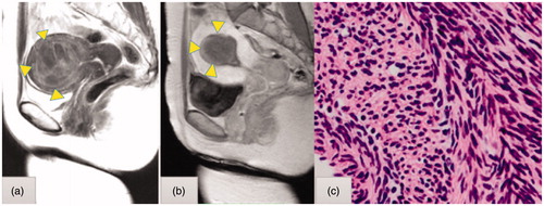
Figure 3. (a) Ultrasonic findings before injection of a dilute vasopressin solution (percutaneous transabdominal cholangiography [PTC] needle). (b) After injection of a dilute vasopressin solution into the space between the myometrium and surface of the leiomyoma using a 21-G PTC needle under transabdominal ultrasound guidance, a hypoechoic lesion into the myometrium was visualized (Δ dilute vasopressin solution).
![Figure 3. (a) Ultrasonic findings before injection of a dilute vasopressin solution (percutaneous transabdominal cholangiography [PTC] needle). (b) After injection of a dilute vasopressin solution into the space between the myometrium and surface of the leiomyoma using a 21-G PTC needle under transabdominal ultrasound guidance, a hypoechoic lesion into the myometrium was visualized (Δ dilute vasopressin solution).](/cms/asset/002ab1eb-d7f4-4628-9986-167f63c9a95a/ihyt_a_1612102_f0003_c.jpg)
Figure 4. Changes in velocity of blood flow before (1) and after (2) injecting a dilute vasopressin solution determined based on the pulsation of the space between the myometrium and surface of the leiomyoma. PI on blood flow in the space changed from 0.94 to 1.40.
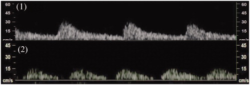
Table 1. Patients characteristics and results.