
Figures & data
Figure 1. Microwave ablation of ectopic secondary hyperparathyroidism nodule in the mandibular area. (A) Hypoechogenic nodule (thin arrow) without blood signal beside the carotid artery (thick arrow) in pre-ablation CDFI ultrasound scan in the 53-year-old male patient. (B) A uniform hyper-enhanced nodule (thin arrow) beside the carotid artery (thick arrow) was displayed in contrast-enhanced ultrasound pre-ablation. (C) Nodule with radioactivity (black arrow) in late phase of MIBI scan. (D) Hyperechoic region inside nodule (thin arrow) beside the carotid artery (thick arrow) during ablation. (E) A non-enhanced area covered the nodule (thin arrow) beside the carotid artery (thick arrow), which suggested complete ablation. (F) Small and iso-echogenic SHPT nodule (thin arrow) beside the carotid artery (thick arrow) was displayed in ultrasound one day after ablation.
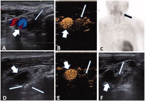
Figure 2. Microwave ablation of ectopic secondary hyperparathyroidism nodule in the suprasternal fossa. (A) A 49-year-old female patient had a hypoechogenic nodule (thin arrow) between the right and left carotid artery (thick arrow) in the suprasternal fossa. (B) MIBI scan (B1) and corresponding CT scan (B2) showing the radioactive nodule located on the suprasternal fossa (cross mark). (C) Ectopic SHPT nodule showing non-uniform hyper-enhancement (thin arrow) in front of the carotid artery (thick arrow) in contrast-enhanced ultrasound pre-ablation. (D) A non-enhanced area covered the nodule (thin arrow) beside the carotid artery (thick arrow), which suggested complete ablation.
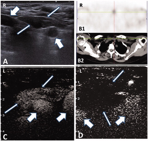
Table 1. Biochemical parameters before and after MWA.