Figures & data
Figure 1. Treitz ligament exploration. This figure illustrates how to explore the peritoneum behind the duodenojejunal flexure, by retracting the first jejunal loop. Panc: pancreas; IMV: inferior mesenteric vein; DJF: duodenojejunal flexure.
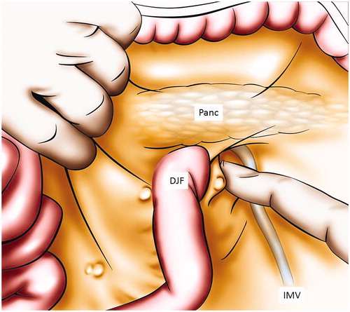
Figure 2. Inter cava caudate peritoneal recess exploration after incision of pars flaccida. This figure illustrates to explore the peritoneum between the vena cava and the caudate lobe by opening the pars flaccida. VC: vena cava; I: liver segment I; LL: liver left lobe; PF: pars flaccida.
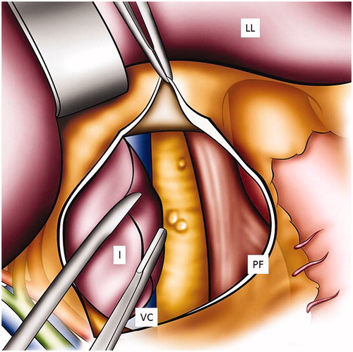
Figure 3. Digital glissonectomy to remove liver surface involvement. This figure illustrates how to expose the peritoneum of the diaphragm and the liver right lobe. A digital peritonectomy of the Glisson capsule is performed. Dia: diaphragm; RL: liver right lobe; GC: Glisson’s capsule; PD: peritonectomy of the diaphragm.
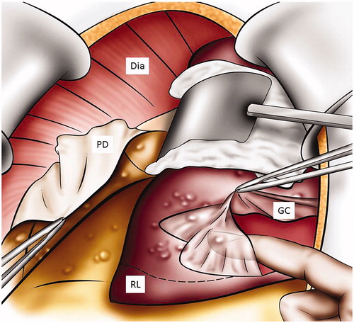
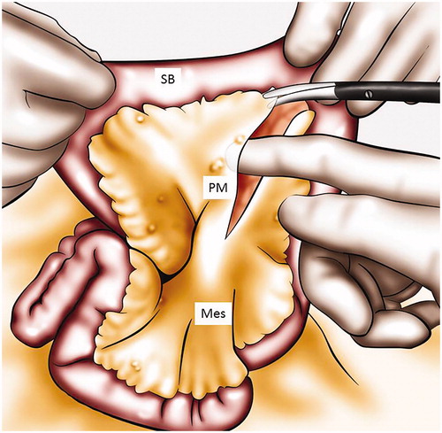
Figure 4. Mesenteric peritonectomy. A peritonectomy of the mesentery is performed. SB: small bowel; Mes: mesentery; PM: peritoneum of the mesentery.