Figures & data
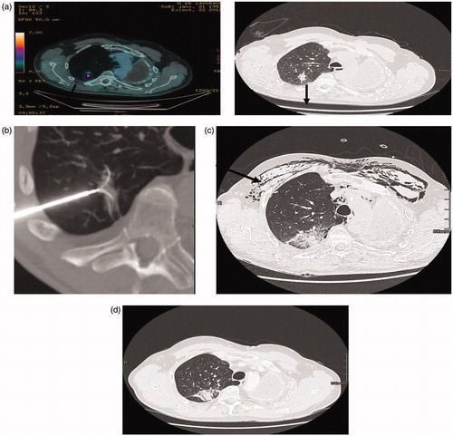
Figure 1. (a) (PET-CT, CT): patient 72 year old, previously treated for an NSCLC by pneumonectomy, 4 years later an adenocarcinoma of the upper right lobe was treated with SBRT recurrence 16 months after SBRT (black arrow). (b) RFA treatment under general anesthesia with a 3.5 cm expandable electrode. (c) CT two days after RFA: the ablation zone encompasses the tumoral volume, subcutaneous emphysema, pneumothorax drained (black arrow). (d) CT 3 months after RFA showing a good result, the ablation zone encompassing the tumor.