
Figures & data
Figure 1. Case of a patient with symptomatic pancreatic adenocarcinoma treated with high intensity focused ultrasound. (a) Magnetic resonance Imaging demonstrating pathologic pancreatic mass. (b) PET/CT scan demonstrating high FDG uptake of the pancreatic tumor. (c) Treatment with US-guided high-intensity focused ultrasound. (d) PET/CT scan after treatment demonstrating lack of FDG uptake in the ablated area.
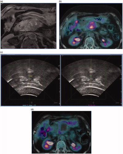
Table 1. Pros and cons of different image-guided ablative modalities for the treatment of pancreatic cancer.