Figures & data
Figure 1. Study Outline: U/L, unilateral; B/L, bilateral; IHC, immunohistochemistry; H&E, hematoxylin & eosin; ACTH, adrenocorticotrophin; HSP, heat shock protein
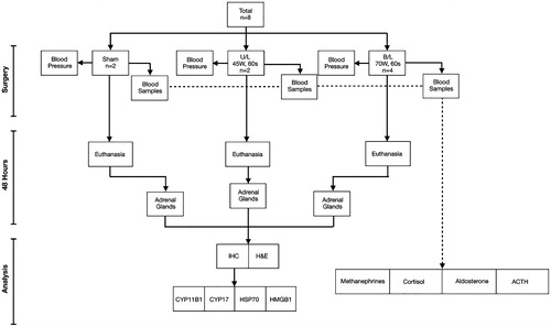
Figure 2. (A) Schema for delivering microwave thermal therapy (MWT) to a benign adenoma in the adrenal target. Micro-wave power is delivered with a side-firing applicator positioned adjacent to the cortex. (B) Temperature profile and estimate of the ablation zone extent for the side-firing ablation probe. (C) Under open surgery in a porcine model, the adrenal gland was exposed by displacing the bowel. (D) The 2.45 GHz side-firing microwave applicator was positioned adjacent to the adrenal gland by direct visualization, and power delivered for 60 s to the cranial pole. (E) Adrenal glands were excised 48 h post-ablation; the region where thermal damage was delivered was identified by visible tissue discoloration, and sections were taken for histopathologic analysis.
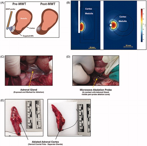
Figure 3. (A) A cross-section of ablated adrenal gland, stained with H&E, demonstrating subtotal ablation of the adrenal gland, with cortical sparing and a delineating transitional zone containing an immune cell infiltrate. (B) Typical features of coagulative necrosis in the adrenal cortex within the so-called ablation zone adjacent to microwave applicator probe placement. (C) Non-targeted adrenal cortex distal to probe placement, which contains zona fasciculata cells of normal morphology, and which are morphologically identical to sham adrenal cortex. The transitional zone between ablated and non-ablated tissue was characterized by an immune infiltrate (D).
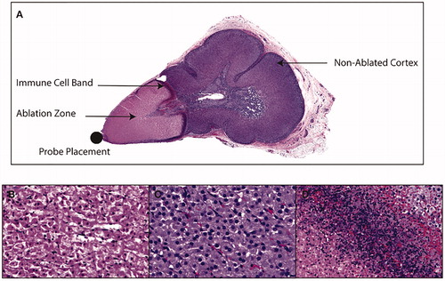
Figure 4. (A) Immunohistochemistry images of expression of the steroidogenic enzyme CYP17 in adrenal gland exposed to high MTA dose. There is an absence of normal cytoplasmic expression of CYP17 in ablated [Ab] tissue as compared to non-ablated tissue [N], and this was sharply delineated by the transitional zone [Tr]. (B) Staining density for CYP17 was markedly reduced within the ablation zone of tissue exposed to high MTA dose (70 W damaged) as compared to sham, low MTA dose (45 W) and non-ablated tissue (70 W undamaged). (C) Immunohistochemistry images of expression of the steroidogenic enzyme CYP11B1 similarly showed disruption of normal cytoplasmic expression in [Ab] tissue as compared to non-ablated tissue [N], and this was also sharply delineated by the transitional zone [Tr]. (D) Staining density tor CYP11B1 was also reduced within the ablation zone of tissue exposed to high MTA dose (70 W damaged) as compared to sham. Low MTA dose (45 W) and non-ablated tissue (70 W undamaged). The immunohistochemical findings are consistent with blood biochemistry which demonstrated appropriate levels of aldosterone (E), cortisol (F) and stimulatory ACTH (G) at 48 h post-operatively. Staining density expressed as percentage (%); ACTH, adrenocorticotrophin; *p < .01 compared with damaged tissue (ANOVA with Tukey's post-hoc test).
![Figure 4. (A) Immunohistochemistry images of expression of the steroidogenic enzyme CYP17 in adrenal gland exposed to high MTA dose. There is an absence of normal cytoplasmic expression of CYP17 in ablated [Ab] tissue as compared to non-ablated tissue [N], and this was sharply delineated by the transitional zone [Tr]. (B) Staining density for CYP17 was markedly reduced within the ablation zone of tissue exposed to high MTA dose (70 W damaged) as compared to sham, low MTA dose (45 W) and non-ablated tissue (70 W undamaged). (C) Immunohistochemistry images of expression of the steroidogenic enzyme CYP11B1 similarly showed disruption of normal cytoplasmic expression in [Ab] tissue as compared to non-ablated tissue [N], and this was also sharply delineated by the transitional zone [Tr]. (D) Staining density tor CYP11B1 was also reduced within the ablation zone of tissue exposed to high MTA dose (70 W damaged) as compared to sham. Low MTA dose (45 W) and non-ablated tissue (70 W undamaged). The immunohistochemical findings are consistent with blood biochemistry which demonstrated appropriate levels of aldosterone (E), cortisol (F) and stimulatory ACTH (G) at 48 h post-operatively. Staining density expressed as percentage (%); ACTH, adrenocorticotrophin; *p < .01 compared with damaged tissue (ANOVA with Tukey's post-hoc test).](/cms/asset/5fa7a075-5bc7-4202-bd0a-45d95c674660/ihyt_a_1650205_f0004_c.jpg)
Figure 5. HSP-70 expression did not differ between non-ablated adrenal cortex exposed to the high MTA dose (A) and sham adrenal cortex (B) at 48 h. A significantly reduced expression was found in ablated cortex as compared to non-ablated, sham and low MTA dose exposed cortex (C). HMGB-1 demonstrated normal nuclear expression, and no difference between non-ablated adrenal cortex was exposed to the high MTA dose (D) and sham adrenal cortex (E). This was confirmed by densitometry measurements of nuclear expression of HMGB-1 (F). Staining density expressed as percentage (%); HSP, heat shock protein; HMGB, high mobility group box. *p < .01 compared with damaged tissue (ANOVA with Tukey's post-hoc test).
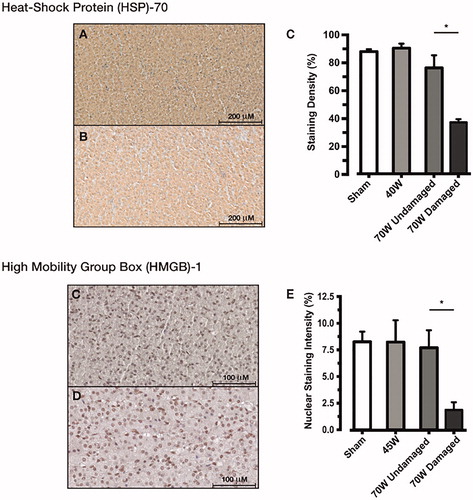
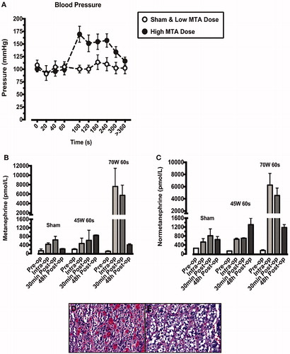
Figure 6. Blood pressure readings were taken throughout each ablation procedure (A). Animals exposed to the higher microwave thermal ablation (MTA) dose (70W for 60s) are represented by closed circles. Sham animals and those exposed to the lower MTA dose (45W for 60s) are represented by open circles. The blood pressure rise demonstrated at 60s in the high MTA dose animals was accompanied by a rise in both metanephrine (B) and normetanephrine (C), indicative of adrenal medullary damage. This was confirmed by H&E staining which demonstrated coagulative necrosis and haemorrhage in adrenal medulla, adjacent to ablated cortex (D). Distal medulla, adjacent to non-targeted cortex retained normal morphology (E).