Figures & data
Table 1. Demographic characteristics of CSP patients with reproductive requirement underwent HIFU followed by USg-D&C (n = 28).
Figure 1. A sagittal view magnetic resonance imaging showed a gestational sac embedding at a previous cesarean section scar (red arrow).
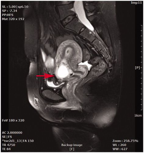
Figure 2. Real-time monitoring ultrasound and contrast enhanced ultrasound images obtained from a patient with CSP before and after HIFU. (A) Pre-HIFU ultrasound image showed a gestational sac (red arrow) embedding in the CSP scar; (B) Real-time ultrasound image showed the significant gray scale change (red arrow) in the embedding area of the CSP scar immediately after HIFU treatment; (C) Pre-HIFU contrast-enhanced ultrasound showed enhancement in myometrium of CSP scar (red arrow) around gestational sac; (D) Post-HIFU contrast-enhanced ultrasound showed no enhancement in myometrium of CSP scar (red arrow).
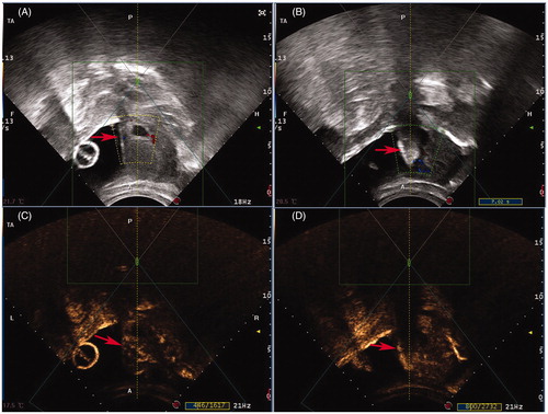
Table 2. Treatment and follow-up outcomes of CSP patients with reproductive requirement underwent HIFU followed by USg-D&C (n = 28).
Table 3. Pregnancy outcomes of CSP patients after HIFU and USg-D&C (n = 23).
Table 4. Delivery outcomes of CSP patients with intrauterine pregnancy after HIFU and USg-D&C (n = 12).
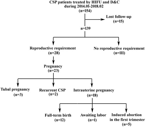
Figure 3. Flow chart of subsequent pregnancy outcomes of CSP patients treated with USgHIFU and USg-D&C.