
Figures & data
Figure 1. Transvaginal ultrasound scan 2 months before MR-HIFU procedure. Uterine fibroid in the anterior wall in direct contact with the endometrium.
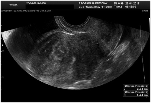
Figure 2. The uterus and the uterine fibroid during MR-HIFU procedure. Thermometry guidance during the thermal ablation.
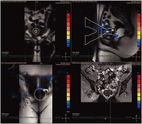
Figure 3. MRI scan of the lower abdomen and pelvis. (A) MR-HIFU qualification MRI scan. (B) MRI scan after the MR-HIFU procedure – visible tumor necrosis.
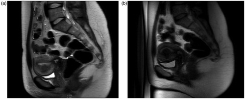
Figure 4. Viable fetus in the uterine cavity – 24th week of the pregnancy. Uterine fibroid located in the anterior wall.
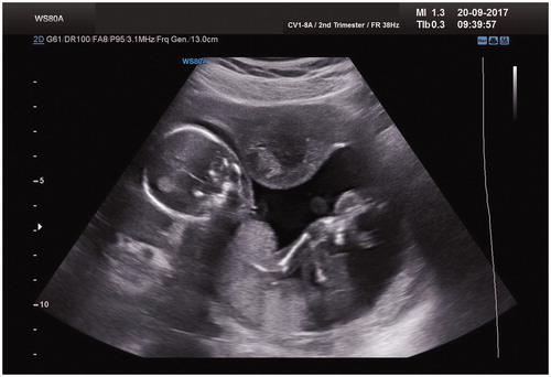
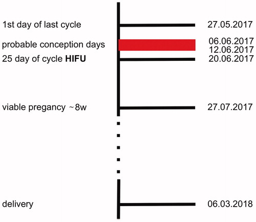
Figure 5. Case report timeline. The MR-HIFU was performed between 8 –12 days after the probable day of conception, according to the last menstruation, and 10 days after conception according to the ultrasound exam.
Data availability statement
The data used to support the findings of this study are available from the corresponding author upon request.