2,169
Views
18
CrossRef citations to date
0
Altmetric
Articles

High-intensity focused ultrasound (HIFU) combined with gonadotropin-releasing hormone analogs (GnRHa) and levonorgestrel-releasing intrauterine system (LNG-IUS) for adenomyosis: a case series with long-term follow up

, , &
Pages 1178-1184 | Received 30 Sep 2018, Accepted 07 Oct 2019, Published online: 03 Dec 2019

Figures & data

Table 1. Baseline characteristics of AD patients.

Figure 1. Two-dimensional transvaginal ultrasonography imaging of uterus by GE Voluson E8 color flow ultrasound machine. (A) Before HIFU treatment, the muscular layer of the anterior wall of the uterus was significantly thickened (30.3 mm, yellow bipolar arrow); with multiple hyperechoic striations and tiny myometrial cysts (white arrows) in the interior. Endometrium was asymmetric. (B) Three months after HIFU treatment, the muscular layer of the anterior wall of the uterus was significantly thinned (21.6 mm, yellow bipolar arrow); with few hyperechoic striations and tiny myometrial cysts (white arrow) in the interior. Endometrium was symmetric.

Figure 1. Two-dimensional transvaginal ultrasonography imaging of uterus by GE Voluson E8 color flow ultrasound machine. (A) Before HIFU treatment, the muscular layer of the anterior wall of the uterus was significantly thickened (30.3 mm, yellow bipolar arrow); with multiple hyperechoic striations and tiny myometrial cysts (white arrows) in the interior. Endometrium was asymmetric. (B) Three months after HIFU treatment, the muscular layer of the anterior wall of the uterus was significantly thinned (21.6 mm, yellow bipolar arrow); with few hyperechoic striations and tiny myometrial cysts (white arrow) in the interior. Endometrium was symmetric.

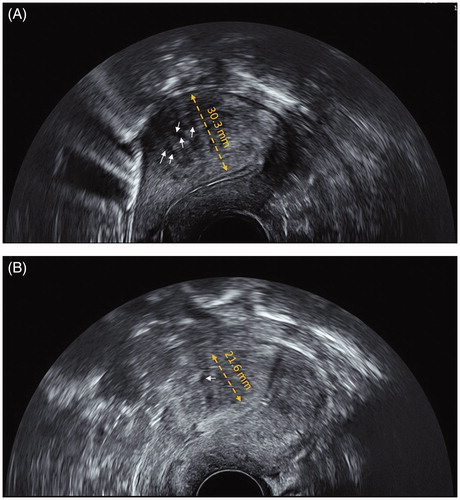
Table 2. Uterine and AD lesions volumes of patients before HIFU and after treatment.

Table 3. Dysmenorrhea score and menstrual flow analysis.

Table 4. Response rate and recurrence rate after treatment.