
Figures & data
Table 1. Inclusion and exclusion criteria.
Figure 1. Patient number 13, 36 years old. (A–C) MR images before HIFU treatment; (D–F) MR images after HIFU treatment. A sagittal T2-weighted image (A) demonstrates anterior wall fibroids (triangle) that are almost hypointense on T2-weighted scans; (C) is a diffusion-weighted image. Fascial swelling has a stripe-like high-intensity signal (long arrow).
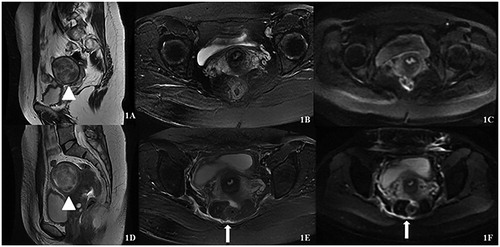
Figure 2. Patient number 125, 39 years old. (A–C) MR images before HIFU treatment; (D–F) MR images after HIFU treatment. A sagittal T2-weighted image (A) demonstrates anterior wall fibroids (triangle) that are almost hypointense on T2-weighted scans; (C) is a diffusion-weighted image. Fascial swelling has a stripe-like high-intensity signal (long arrow).
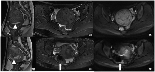
Table 2. Univariate analysis to evaluate the relationship between the endopelvic fascial swelling and the features of fibroids.
Table 3. Univariate analysis to evaluate the relationship between the endopelvic fascial swelling and ultrasound ablation parameters.
Table 4. Multivariable binary logistic regression analysis to evaluate the correlation of fascial swelling with the significant factors of univariate analysis.
Table 5. Correlation between the location of the fibroid and the degree of endopelvic fascial swelling.
Table 6. Correlation among the quantitive factors (TD, sonication time, EEF and the distance from the fibroid to the sacrum) and the degree of endopelvic fascial swelling.
Table 7. Summary of postoperative adverse events.
Data availability statement
The data that support the findings of this study are available from the corresponding author Zhang upon reasonable request.