
Figures & data
Table 1. Baseline characteristics of patients and lesions.
Figure 1. A 66-year-old male. (A) Contrast-enhanced MRI indicated a recurrent hepatocellular carcinoma located in segment 4/8 with a maximum diameter of 17 mm. (B) The nodule was partially visible and partially affected by the pulmonary gas. (C and D) The nodule became completely visible after one-lung ventilation and contrast-enhanced ultrasound confirmed the target location. (E) Thermal ablation was performed and immediate contrast-enhanced ultrasound demonstrated that the avascular zone covered the index tumor completely. (F) Contrast-enhanced MRI one month after the ablation procedure confirmed the technical efficacy.
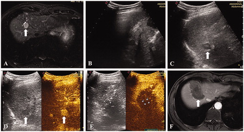
Figure 2. A 48-year-old male. (A) Contrast enhanced MRI indicated a recurrent hepatocellular carcinoma of 12 mm in segment 7. (B) The nodule was completely invisible because of the pulmonary gas. (C) The nodule became completely visible after one-lung ventilation. (D) Thermal ablation was performed and immediate contrast-enhanced ultrasound demonstrated no hyper-enhancement of the tumor. (E) Contrast-enhanced MRI one month after the ablation procedure confirmed the technical efficacy.
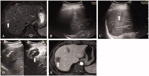
Table 2. Visibility scores before and after one-lung ventilation.