
Figures & data
Figure 1. CONSORT diagram for the study.
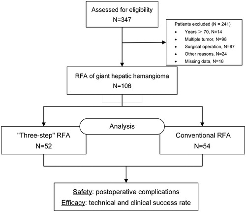
Figure 2. Percutaneous ultrasound-guided ‘three-step’ radiofrequency ablation of hepatic hemangioma located in the right lobe. (A) Contrast-enhanced ultrasonography is used to locate the tumor (the tumor was 7.66 cm in diameter) and its feeding artery. (B) The first step is to ablate the feeding artery. (C) The second step is to aspirate blood from the tumor (the diameter of the tumor after the blood was drawn was 4.11 cm). (D) The third step is to ablate the lesion.
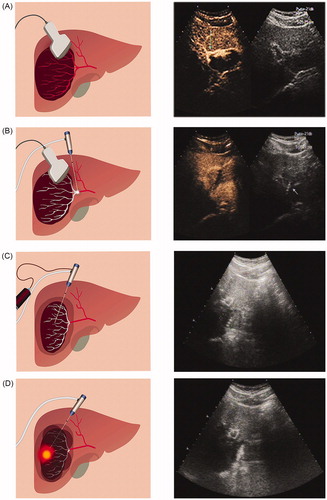
Table 1. Patient and tumor characteristics between the two study groups.
Table 2. Intraoperative information and postoperative outcomes.
Figure 3. Comparison of preoperative and postoperative liver function, renal function and hemoglobin indexes.
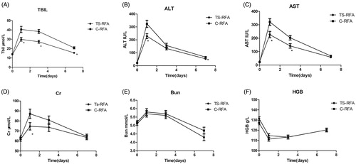
Table 3. Postoperative morbidity.