1,601
Views
15
CrossRef citations to date
0
Altmetric
Article

Clinical efficacy and mechanism for focused ultrasound (FUS) in the management of cervical intraepithelial neoplasia 1 (CIN1)

, , , , , & show all
Pages 339-345 | Received 20 Oct 2019, Accepted 24 Mar 2020, Published online: 09 Apr 2020

Figures & data

Table 1. Characteristics of the enrolled patients with CIN.

Figure 1. (a) The cervical appearance of a patient under colposcopy before focused ultrasound treatment. Acetic acid staining showed white plaque on the outside of the cervix (CIN1 for cervical biopsy. (b) The cervical appearance of the same patient under colposcopy at 3 months after treatment. The cervix is completely back to normal. (c) the expressions of P16 in cervical tissue were strongly positive and darkly staining (immunostaining, 400 × magnification) before treatment; 1d. 3 months after treatment, the expressions of P16 were weakly positive with light staining, and the number of cells of positive expressions had reduced; 1e. the expressions of Ki-67 in cervical tissue were strongly positive and darkly staining (immunostaining, 400x magnification) before treatment; 1f. 3 months after treatment, the expressions of Ki-67 were weakly positive with light staining, and the number of cells of positive expressions had reduced; 1g. the expressions of Fas at lower 1/3 of the cervical epithelium were weakly positive with light staining, and the number of cells of positive expressions was less (immunostaining, 400x magnification) before treatment; 1h. 3 months after treatment, the expressions of Fas were strongly positive and with darker staining, and the number of cells with positive expression had increased compared to before treatment.

Figure 1. (a) The cervical appearance of a patient under colposcopy before focused ultrasound treatment. Acetic acid staining showed white plaque on the outside of the cervix (CIN1 for cervical biopsy. (b) The cervical appearance of the same patient under colposcopy at 3 months after treatment. The cervix is completely back to normal. (c) the expressions of P16 in cervical tissue were strongly positive and darkly staining (immunostaining, 400 × magnification) before treatment; 1d. 3 months after treatment, the expressions of P16 were weakly positive with light staining, and the number of cells of positive expressions had reduced; 1e. the expressions of Ki-67 in cervical tissue were strongly positive and darkly staining (immunostaining, 400x magnification) before treatment; 1f. 3 months after treatment, the expressions of Ki-67 were weakly positive with light staining, and the number of cells of positive expressions had reduced; 1g. the expressions of Fas at lower 1/3 of the cervical epithelium were weakly positive with light staining, and the number of cells of positive expressions was less (immunostaining, 400x magnification) before treatment; 1h. 3 months after treatment, the expressions of Fas were strongly positive and with darker staining, and the number of cells with positive expression had increased compared to before treatment.

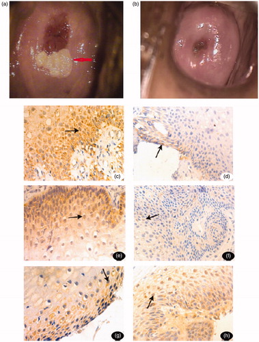
Table 2. Changes in symptoms, cytology and biopsy before and after FUS therapy.

Table 3. The expression of P16, Ki-67 and Fas before and after treatment (n = 30).