
Figures & data
Table 1. patient demographic and laboratory details.
Figure 1. Single-fiber laser ablation for primary hyperparathyroidism. (A) A 69-year-old female presented with severe osteoporosis, nephrocalcinosis and elevated parathormone levels. A 1.6 × 0.6 cm adenoma was identified behind the lower pole of the thyroid. (B) Contrast enhanced ultrasound confirms marked homogeneous vascularity. (C) Gray scale ultrasonography demonstrates insertion of a single fiber (central white line) along the longitudinal axis of the gland. Normalization of parathormone was noted at 6 months. (D) Two-year follow-up shows a marked reduction in the gland size (0.7 × 0.4 cm) with elimination of vascularity at contrast enhanced ultrasound (yellow circle).
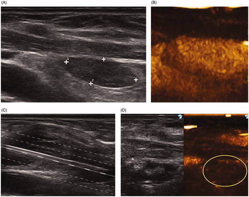
Figure 2. Dual-fiber laser ablation for primary hyperparathyroidism. (A) A 65-year-old female presented with increasing bone pain and arthralgia, increased parathormone levels and hypercalcemia. A 2.0 × 1.3 cm adenoma was identified at gray scale ultrasound caudal to the thyroid. (B) Contrast enhanced ultrasound demonstrated a heterogeneously enhancing nodule. (C) Baseline Tc-99 Sestamibi is highly positive (red color). (D) Intraprocedural gray scale ultrasound demonstrated the placement of two laser fibers spaced 1 cm apart inserted into the gland. (E) Increased echogenicity is identified surrounding the fibers during the active application of laser energy. Normalization of parathormone and calcium levels occurred by 6 months. (F) Two-year follow-up gray scale and contrast enhanced ultrasound demonstrate a non-enhancing 0.9 × 0.7 cm gland. (G) Tc-99 Sestamibi shows no evidence for increased uptake.
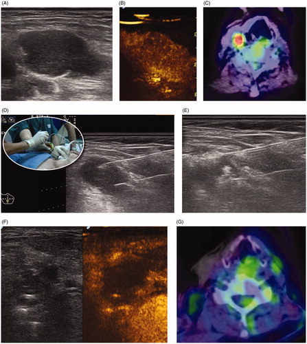
Table 2. The changes in serum PTH and albumin- corrected calcium levels before ablation and at each follow-up period.