Figures & data
Table 1. Baseline characteristics of the 84 enrolled patients.
Table 2. Treatment results of uterine fibroids treated by HIFU.
Figure 1. AMH values of the patients younger than 35 years of age.
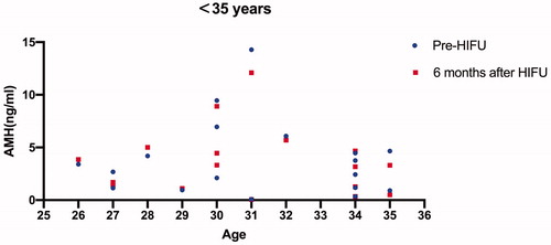
Table 3. AMH levels in different age groups assessed before and 6 months after HIFU.
Figure 2. AMH values of the patients between 36 and 40 years of age.
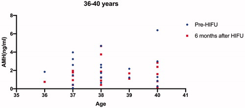
Figure 3. AMH values of the patients between 41 and 45 years of age.
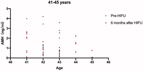
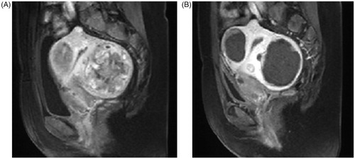
Figure 4. A sagittal plane of contrast enhanced MRI obtained from a 43-year-old patient with multiple uterine fibroids before and 1 day after HIFU treatment. (A) pre-HIFU image showed multiple uterine fibroids with poor to medium blood supply; (B) post-HIFU image showed the multiple uterine fibroids were completely ablated without damaging to the normal structures of the uterus.