
Figures & data
Figure 1. A 47-year-old woman who had undergone a total thyroidectomy 2 years ago for papillary thyroid cancer underwent microwave ablation (MWA) of cervical metastatic lymph nodes (LN). A, Pre-MWA, B-mode ultrasonography (US) showed hypoechoic LN (arrow); B, Fine-needle aspiration biopsy (FNAB) process of the LN (arrow); C, Pre-MWA, the contrast-enhanced US (CEUS) showed uneven and highly enhanced patterns (arrow); D, Spacer fluid (arrow) was injected to surround the LN; E, Hyperechoic (arrow) pattern in the LN during MWA; F, Post-MWA, the CEUS showed no enhancement (arrow) in LN.
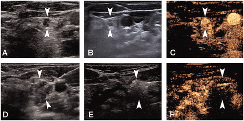
Table 1. Clinical characteristics of the study population.
Table 2. Characteristics of cervical metastatic lymph nodes.
Table 3. Changes in cervical metastatic lymph nodes post-ablation at each follow-up.