Figures & data
Figure 1. LITT operating room workflow. (A) At our center, bone fiducials and OArm CT are used for frameless registration. The robot is used for placement of the LITT guidance bolt. (B) Intra-ablation MR thermography provides online feedback demonstrating the extent of ablation. (C) Intra-operative post-ablation post-contrast T1-weighted MR showing ablation relative to MR thermography ablation volume.
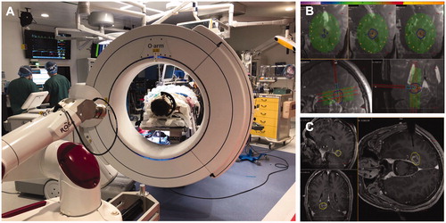
Figure 2. Stereotactic LITT completion callosotomy in a patient with previous callosotomy. (A and B) Preoperative T1 axial (A) and sagittal (B) imaging demonstrating residual anterior corpus callosum and splenium of corpus callosum, which are targets of the planned ablation. (C) Intra-ablation MR thermography demonstrating ablation of the residural genu and splenium of the corpus callosum. (D and E) Postoperative axial (D) and sagittal (E) DWI MR images demonstrating complete callosotomy.
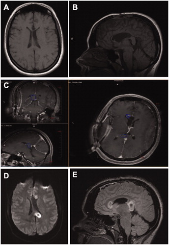
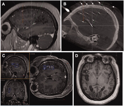
Figure 3. sEEG-guided ablation of insulo-opercular epilepsy. (A) sEEG trajectory planning; (B) post-sEEG implantation CT demonstrating electrode contact corresponding to the seizure onset zone; (C) intra-ablation MR thermography demonstrating ablation of the insulo-opercular target. (D) Postoperative (3 months) T1-weighted MR image showing insulo-opercular lesion.