7,537
Views
54
CrossRef citations to date
0
Altmetric
Reviews

Hyperthermia treatment advances for brain tumors

, , , , , & show all
Pages 3-19 | Received 22 Feb 2020, Accepted 16 May 2020, Published online: 16 Jul 2020

Figures & data

Figure 1. Brain tumor illustration demonstrating laser interstitial thermal therapy (LITT). (A) Schematic representation of intratumoral placement of laser catheter and brain tumor ablation. (B) Schematic representation of brain tumor ablation demonstrating post-LITT contrast enhancement consistent with LITT related blood brain barrier (BBB) disruption and LITT related perifocal edema.

Figure 1. Brain tumor illustration demonstrating laser interstitial thermal therapy (LITT). (A) Schematic representation of intratumoral placement of laser catheter and brain tumor ablation. (B) Schematic representation of brain tumor ablation demonstrating post-LITT contrast enhancement consistent with LITT related blood brain barrier (BBB) disruption and LITT related perifocal edema.

Figure 2. Schematic representation of magnetic hyperthermia therapy in the brain. (A) Alternating magnetic field is applied to the patient after local administration of magnetic nanoparticles (black spheres), generating highly localized hyperthermia. (B) Heat is produced via hysteresis losses and Brownian relaxation (a process in which frictional heating is generated by the physical rotation of the magnetic particle).

Figure 2. Schematic representation of magnetic hyperthermia therapy in the brain. (A) Alternating magnetic field is applied to the patient after local administration of magnetic nanoparticles (black spheres), generating highly localized hyperthermia. (B) Heat is produced via hysteresis losses and Brownian relaxation (a process in which frictional heating is generated by the physical rotation of the magnetic particle).

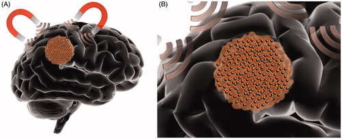
Table 1. Nanoparticle constructs utilized for magnetic hyperthermia therapy.

Figure 3. Illustration of brain tumor nanoparticle-mediated photothermal therapy (PTT). (A) Schematic representation of local NIR light application to a brain tumor after photothermal agents (PTAs) administration (black spheres). (B) Schematic illustration demonstrating NIR light excitation of PTAs within the brain tumor. PTAs absorb NIR light and emit thermal energy acting as local heat sources.

Figure 3. Illustration of brain tumor nanoparticle-mediated photothermal therapy (PTT). (A) Schematic representation of local NIR light application to a brain tumor after photothermal agents (PTAs) administration (black spheres). (B) Schematic illustration demonstrating NIR light excitation of PTAs within the brain tumor. PTAs absorb NIR light and emit thermal energy acting as local heat sources.

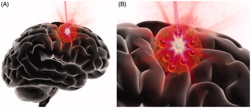
Table 2. Nanoparticle constructs utilized for photothermal therapy.

Figure 4. Schematic illustration summarizing some of the less common HT modalities that have been employed for brain tumor treatment. HT for brain tumors has been performed with ultrasound (US), radiofrequency (RF) and microwaves (MW).

Figure 4. Schematic illustration summarizing some of the less common HT modalities that have been employed for brain tumor treatment. HT for brain tumors has been performed with ultrasound (US), radiofrequency (RF) and microwaves (MW).

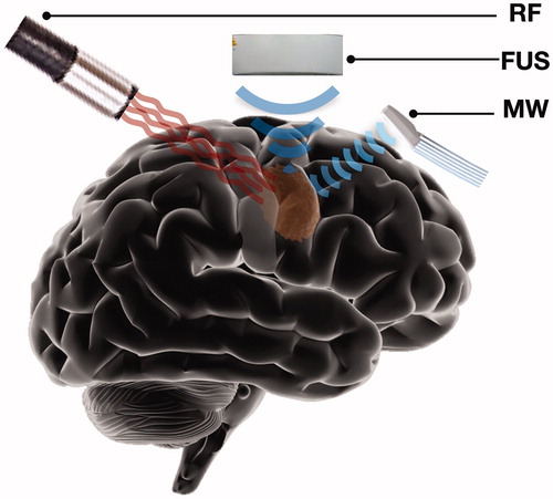
Table 3. Understudied HT modalities for the treatment of brain tumors.