
Figures & data
Figure 1. Contrast-enhanced CT images for a representative case involving a 49-year-old woman with a benign follicular nodule in the left thyroid gland prior to treatment. CT-based measurements of the (a) tracheal deviation distance, (b) ratio of anteroposterior/transverse diameter of the trachea, and (c) anterior neck angle.
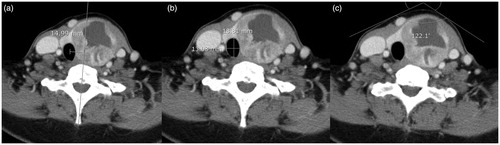
Table 1. Baseline characteristics of patients who underwent radiofrequency ablation for benign thyroid nodules.
Figure 2. Contrast-enhanced axial (a, b) and coronal (c, d) CT images for a representative case involving a 32-year-old woman with benign nodular hyperplasia in the left thyroid gland. Volumetric segmentations of the thyroid glands are shown in red while cross-sectional segmentation of the trachea is shown in blue.
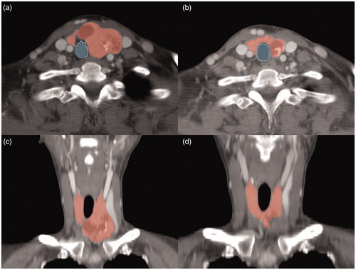
Figure 3. Mean volume reduction rates (with standard errors of means) measured by CT and US according to the number of radiofrequency ablation (RFA) sessions; no significant differences in volume reduction rates was observed according to the number of RFA sessions.
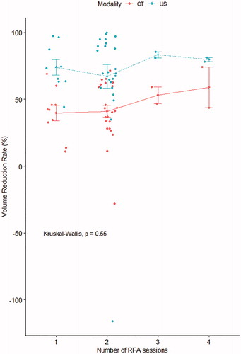
Figure 4. Contrast-enhanced CT images obtained (a) before and (b) after two radiofrequency ablation sessions (interscan interval: 4.5 years) for a 50-year-old woman with a benign nodule in the right thyroid gland. The volume reduction rate was high (65.5%). Normalization of adjacent structures, including an increase in the tracheal area (4 mm2) and anterior neck angle (20°), a decrease in the ratio of anteroposterior/transverse diameter of the trachea (0.31), and a reduction in midline deviation of the trachea (2.3 mm), could be observed.
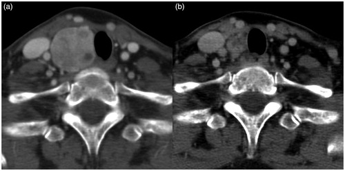
Table 2. Comparison of CT-based measurements obtained before and after radiofrequency ablations for benign thyroid nodules.