Figures & data
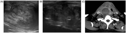
Figure 1. Consort diagram of the study.
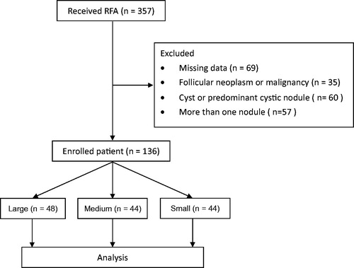
Table 1. Patient and nodule characteristics based on nodule volume.
Figure 2. Changes in nodular volume and VRR of the three volume groups of BTN at each follow-up. Changes in nodular volume (A) and VRR (B) of the different echogenicity groups of BTN at each follow-up. BTN: benign thyroid nodule; VRR: volume reduction ratio.
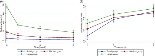
Table 2. Outcomes based on nodule volumea.
Figure 3. Goiter rupture before and after RFA. Ultrasound and CT obtained after nodular rupture of the patient with previous partial thyroidectomy history and received thyroid RFA. (A) Incomplete anterior thyroid capsules surrounding the thyroid nodule noted before RFA. (B)The incomplete capsule site is afterward the nodular rupture site on sonography. (C) Post-contrast CT scan shows a ruptured nodule into the right anterior neck. RFA: radiofrequency ablation.