Figures & data
Figure 1. Transverse view of MR images obtained before and 1 day after HIFU treatment. (A). A pre-HIFU T2 weighted image showed multiple nodules with the largest one at a 6 cm protruding into the uterine cavity; (B). A post-HIFU T2 weighted image showed the signal intensity of the largest fibroid decreased; (C). A pre-HIFU contrast MR image showed hyperenhancement of the fibroids; (D). A post-HIFU contrast MR image showed no perfusion in the largest fibroid. The largest fibroid was completely ablated.
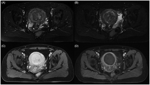
Figure 2. Intraoperative photography. (A) Multinodular solid mass that originated from serosal surface in the pelvis (left). (B) Small lesions located on the surface of the left lobe of the liver (right).
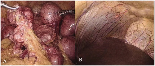
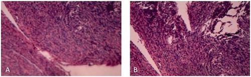
Figure 3. Microscopic examination revealed smooth muscle proliferation (hematoxylin and eosin 10 × 10).