
Figures & data
Figure 1. A 22-year-old male patient with PTC was found to have metastatic lymph nodes in area VI of the right neck (Ultrasound image obtained from head side of patients). The lymph node was located between the common carotid artery and the trachea. The size of lymph nodes was about 1.2cmx1.2 cm.
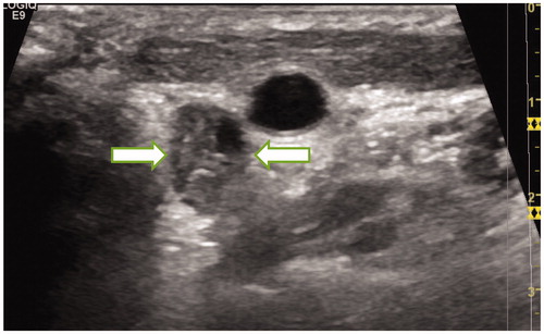
Figure 2. The CEUS of the metastatic lymph nodes in the above patients showed a high enhancement mode.
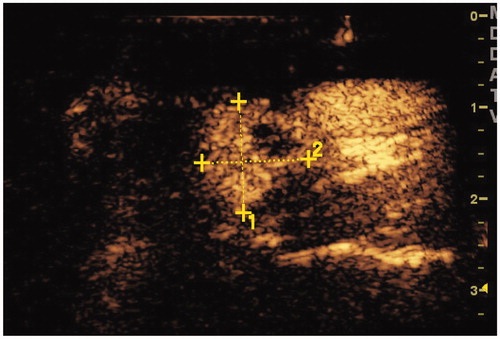
Figure 3. The flowchart of patient selection process.
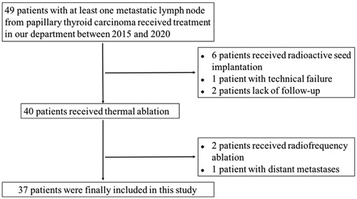
Figure 4. Normal saline was injected between the targeted lymph nodes and the trachea to isolate them.
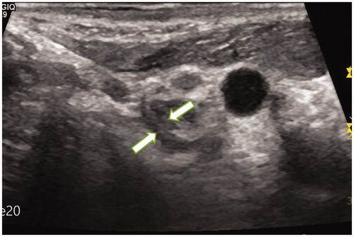
Figure 5. The patient received MWA ablation of metastatic lymph nodes in July 2016. Ultrasound monitoring showed that some lymph nodes were covered by microbubbles.
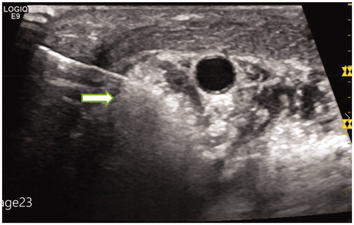
Figure 6. On the second day after ablation, CEUS showed that the ablated lymph nodes had no enhancement (ultrasound image obtained from foot side of patients).
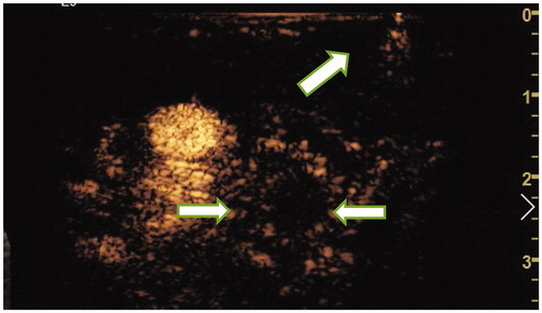
Figure 7. The patient was last reexamined in January 2020. Ultrasound images showed that the size of the ablated lymph nodes was significantly reduced. The size of lymph nodes was about 0.61cmx0.57 cm.
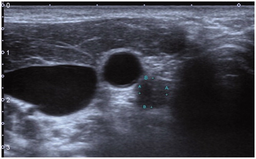
Table 1. Characteristics of the targeted lymph nodes before and after MWA.