Figures & data
Figure 1. Flowchart of patient enrollment.
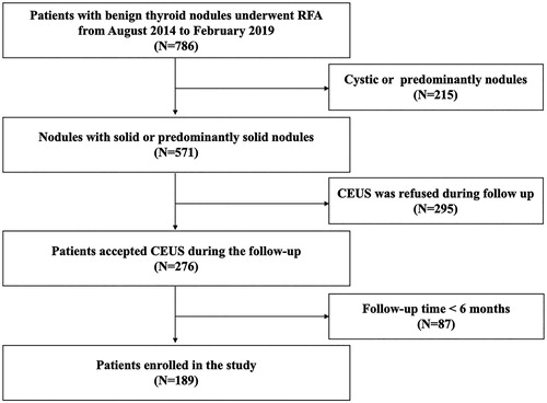
Table 1. Clinical characteristics of patients before RFA.
Figure 2. The changes of VRR at each follow-up point after RFA.
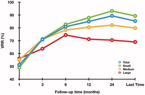
Table 2. Changes of VRR in the subgroups at each follow-up period after RFA.
Figure 3. The changes of Vt, Va and Vv in each subgroup at each follow-up point after RFA (A: small subgroup; B: medium subgroup; C: large subgroup).
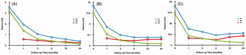
Table 3. Changes of Vt, Va and Vv in the subgroups at each follow-up period after RFA.
Table 4. Univariate and multivariate logistic regression analyses for nodule regrowth.
Figure 4. The ROC curve for RVR to predict regrowth.
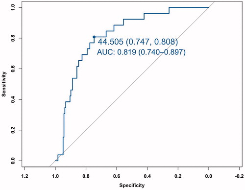
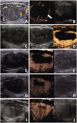
Figure 5. The conventional US and CEUS images of a 48-year-old male with a benign thyroid nodule. (A) Before RFA, a solid thyroid nodule located in the right thyroid lobe with an initial volume of 22.27 ml. Color Doppler showed the vascularity was grade 3. (B) CEUS performed immediately after RFA showed a lack of enhancement on the treated area(arrow). (C, D) At one months after RFA, conventional US showed the treated nodule began to shrink and total volume(Vt) was 5.28ml. CEUS showed the treated nodule was divided into the centrally non-enhancement area(ablated volume, Va) and peripheral iso-enhancement area(vital volume, Vv). Va was 2.59ml and Vv was 2.69ml. RVR was 50.95%. (E, F) At 3 months after RFA, conventional US showed Vt was 3.48ml and the echogenicity of nodule was heterogeneous. Va decreased to 1.11ml on CEUS and Vv was 2.37ml. (G, H) At 6 months after RFA, conventional US and CEUS showed Vt decreased to 2.89 ml and Va decreased to 0.27ml, respectively. However, Vv began to enlarge to 2.62ml.(I, J) At 12 months after RFA, conventional US showed nodule regrowth and Vt was 4.34ml. Va decreased to 0.20ml on CEUS and thus Vv increase also occurred and Vv was 4.13ml.