
Figures & data
Figure 1. (a) Axial pretreatment post-contrast CT-image of a 72-year-old woman, showing a metastasis in the VI hepatic segment originating from a leiomyosarcoma. (b) Selective diagnostic hepatic angiography showing the right hepatic artery and the feeding vessels to the liver metastasis. (c) US image performed during the MWA procedure showing the antenna correctly inserted into the liver tumor. (d) The post-embolization angiography shows a contrast leak, successfully treated using microcoils. (e) The selective diagnostic hepatic angiography shows the embolization with coils of the bleeding vessel. (f) 1-month axial CT-image control showing no residual tumor.
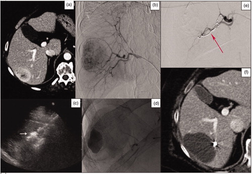
Figure 2. (a) Axial pretreatment post-contrast CT exam of a 54-year-old woman, showing a polilobated hypoenhancing colon cancer metastasis in segment IV. (b) Superselective angiography through the right femoral artery. A subsequent post-contrast ultrasound scan confirmed the artery network; embolization was performed by administering 75 μm Embozene particles. (c) Ultrasonographic monitoring of the subsequent MWA treatment. (d) Post-treatment axial CT-image control, showing a large area of necrosis. (e) PET-CT performed at 1 year showing no pathologic uptake in the treated area. (f) MRI-image control at 1-year and post-contrast CT scan at 2 years showing a decrease in the necrotic area.
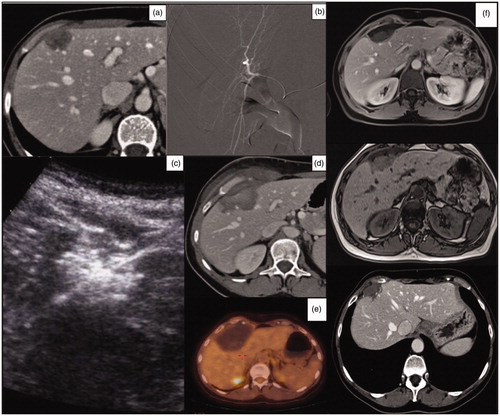
Figure 3. (a) Axial venous phase contrast-enhanced CT scan image of a 76-year-old woman, showing a gastric neuroendocrine metastasis in the V-VI hepatic segment. (b) Selective diagnostic angiography of the hepatic artery showing the tumor. (c) Post-TAE selective diagnostic angiography showing the complete devascularisation of the tumor. (d) Axial venous phase post-contrast CT-image showing the embolized tumor with a necrotic enlarged area.
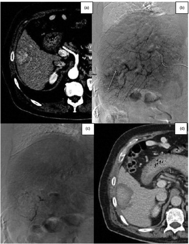
Figure 4. (a) Axial CT-image of a 74-year-old woman, showing a breast metastasis in the VI hepatic segment. (b, c) Selective diagnostic hepatic angiography, showing the hepatic vascularization of the right lobe and the embolization of the vessels feeding the liver metastasis. (d) US-monitoring of the subsequent MWA procedure. (e, f) US contrast-image performed 6 months after the TAE-RFA procedure, showing a large area of necrosis and no residual tumor.
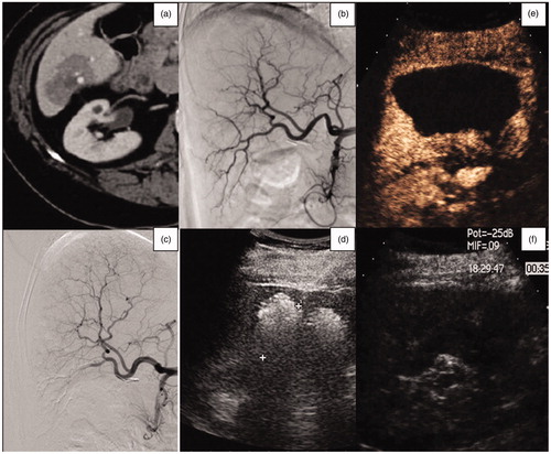
Figure 5. Comparison of the ablation volumes obtained using the combined treatment (red bars), ablation volumes which would have been obtained using stand-alone MWA (blue bars), and the pretreatment volumes of the nodules (yellow bars).
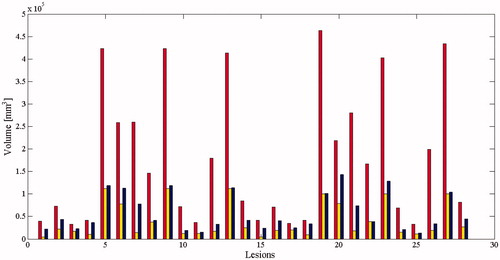
Table 1. Influence of technical and anatomic parameters on the necrotic area dimensions.