
Figures & data
Figure 1. Flow chart of the study.
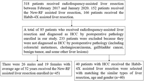
Table 1. Baseline characteristics of enrolled patients.
Figure 2. The electrode was inserted along the marked line to coagulate the liver tissue.
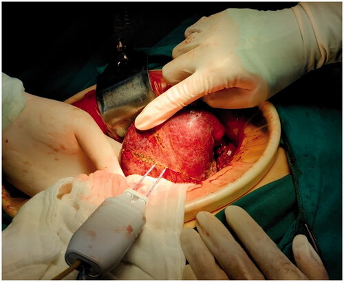
Figure 3. Vascular forceps or tissue scissors were used to excise the liver along the ‘no blood circulation’ tissue plane.
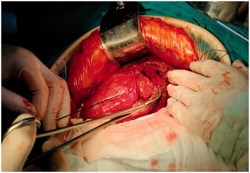
Figure 4. The process obeyed the sequence of ‘coagulation to excision’, which allowed us to change the direction during the liver excision according to the position, size and shape of the tumor.
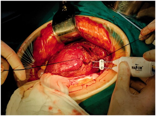
Table 2. Intraoperative data of enrolled patients.
Table 3. Lab examinations of patients after surgery.
Table 4. Complications of patients after surgery.