2,561
Views
5
CrossRef citations to date
0
Altmetric
Articles

High-intensity focused ultrasound ablation in the treatment of recurrent ovary cancer and metastatic pelvic tumors: a feasibility study

, , , , , , & show all
Pages 282-287 | Received 15 Jan 2021, Accepted 08 Feb 2021, Published online: 22 Feb 2021

Figures & data

Figure 1. The real-time ultrasound image obtained from a 46-year-old patient with metastatic pelvic tumor from endometrial cancer. (A). A pre-HIFU ultrasound image showed a recurrent lesion of 112 cm × 72cm × 71cm, with mixed echoic (arrow); (B). During HIFU treatment, a significant gray scale changed area was observed in the pelvic lesion (arrow); (C) ultrasound image obtained at 1301 s of sonication showed the gray scale changed area covered the pelvic lesion (arrow).

Figure 1. The real-time ultrasound image obtained from a 46-year-old patient with metastatic pelvic tumor from endometrial cancer. (A). A pre-HIFU ultrasound image showed a recurrent lesion of 112 cm × 72cm × 71cm, with mixed echoic (arrow); (B). During HIFU treatment, a significant gray scale changed area was observed in the pelvic lesion (arrow); (C) ultrasound image obtained at 1301 s of sonication showed the gray scale changed area covered the pelvic lesion (arrow).

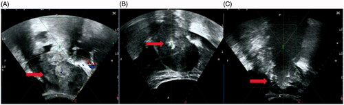
Table 1. Baseline characteristics of patients with metastatic pelvic tumors.

Figure 2. Pre- and post-HIFU MRI images obtained from a 71-year-old patient with pelvic recurrence and metastasis from ovarian cancer. (A). Pre-HIFU MRI image showed a lesion adjacent to the bladder. The size of the lesion was 33 cm × 42cm × 45cm with significantly enhancement (red arrow). (B). One month after HIFU, contrast-enhanced MR showed a non-enhanced area of 30 cm × 33cm × 36cm (arrow).

Figure 2. Pre- and post-HIFU MRI images obtained from a 71-year-old patient with pelvic recurrence and metastasis from ovarian cancer. (A). Pre-HIFU MRI image showed a lesion adjacent to the bladder. The size of the lesion was 33 cm × 42cm × 45cm with significantly enhancement (red arrow). (B). One month after HIFU, contrast-enhanced MR showed a non-enhanced area of 30 cm × 33cm × 36cm (arrow).

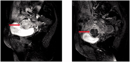
Table 2. HIFU treatment results for metastatic pelvic tumors.

Table 3. Adverse effects evaluation of HIFU for metastatic pelvic tumors.

Table 4. Comparison of pain scores during follow-up period.