
Figures & data
Figure 1. (a, b) Three electrodes with 4-cm tips were arranged in a linear manner using a linear adaptor.
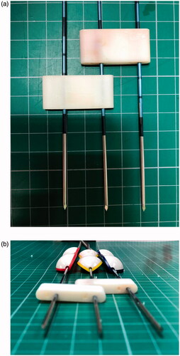
Figure 2. Electrodes were inserted into the pre-determined resection plane in a linear arrangement to conduct ablation.
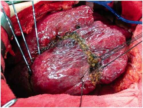
Figure 3. (a, b) After the completion of ablation, transection of the liver parenchyma was performed using a scalpel.
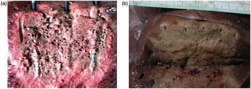
Table 1. Experimental groups and mode (DS or SS), electrode interval (2 cm or 3 cm) and ablation time (1.5, 2 or 3 min).
Table 2. Perioperative ablation data and experimental outcomes.