Figures & data
Table 1. The characteristics of the patients and tumors.
Figure 1. Contrast-enhanced MRI of three types of desmoid tumors (DTs). Before HIFU ablation, the areas of significant enhancement (white arrow) were shown in extra-abdominal DT (A), abdominal wall DT (B) and intra-abdominal DT (C). One week after HIFU ablation, non-perfused areas (red arrow) were observed within the extra-abdominal DT (A1), abdominal wall DT (B1) and intra-abdominal DT (C1), and the areas of peripheral enhancement representing residual tumors (blue arrow) were observed in the extra-abdominal DT (A1) and intra-abdominal DT (C1).
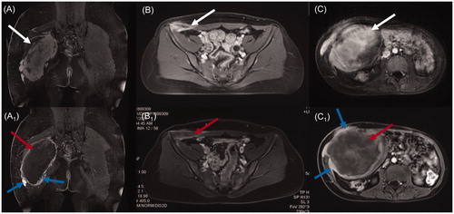
Table 2. The treatment parameters and HIFU ablation results.
Table 3. The types and incidences of adverse events in 188 sessions of HIFU ablation.
Table 4. Comparison of the incidences of adverse events between patients with a single session of HIFU ablation and those with multiple sessions of HIFU ablation.
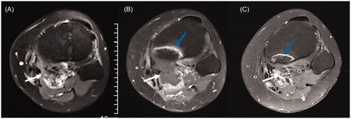
Figure 2. Contrast-enhanced axial MRI obtained from a 26-year-old woman with recurrent DT in the popliteal fossa before and after HIFU ablation. (A) Before HIFU ablation, the DT showed significant enhancement (white arrow), and the tibia showed no abnormal signal. (B) Three months after HIFU ablation, the DT without enhancement shrank (white arrow), and the tibia adjacent to the DT showed banded hyperintensity (blue arrow). (C) Fifteen months after HIFU ablation, the DT disappeared (white arrow), and the area of hyperintensity in the tibia (blue arrow) shrank.