
Figures & data
Figure 1. Follow chart of this study. PTC: papillary thyroid carcinoma; PTMC: papillary thyroid microcarcinoma; RFA: radiofrequency ablation.
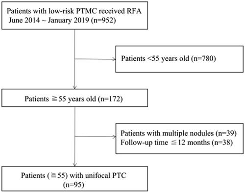
Table 1. Demographics and characteristics of the nodules.
Table 2. RFA information of the 95 nodules.
Table 3. Complications after RFA.
Figure 2. A 60-year old woman with a papillary thyroid carcinoma in the middle of the left thyroid lobe (A) A hypoechoic nodule (white arrow) with an irregular margin was detected in the middle of the left thyroid lobe. (B) Immediately after radiofrequency ablation (RFA), a non-enhanced area measuring 1.4 × 1.4 × 1.4 cm was observed in the ablation zone. (C–E) Follow-up images showed that the ablation zone decreased gradually to 1.1 × 1.1 × 0.8, 0.9 × 1.0 × 0.8, and 0.6 × 0.7 × 0.5 cm at 1, 3 and 6 months, respectively. (F) At 12 months after RFA, the ablation area had disappeared on ultrasonography.
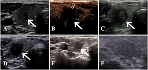
Figure 3. Tumor disappearance time curve after ablation. 46.3% of tumors completely disappeared within 12 months during their follow-up period.
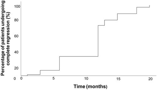
Table 4. Volume changes of the nodules in the 95 patients.