Figures & data
Table 1. The data of patients with broad ligament fibroids and ultrasound-guided HIFU treatment results.
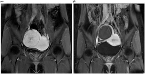
Figure 1. Contrast enhanced-MRI images of a 36-year-old woman with a right broad ligament fibroid. These showed a moderately vascular fibroid before treatment (A) and more than 90% NPV ratio immediately after HIFU ablation (B).
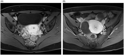
Figure 2. A 42-year-old woman with a left broad ligament uterine fibroid and a small submucous fibroid. The fibroid was hypo-intense on T2WI MRI (A) and hypervascular on CE-MRI before treatment (B). There was localized edema and swelling of abdominal muscles (white arrow) (C) and the NPV ratio of the treated fibroid was 91.3% immediately after treatment (D). No abnormal MRI finding related to focused ultrasound ablation was observed (E) and the fibroid volume decreased by 55.5% at 6-month follow-up (F).
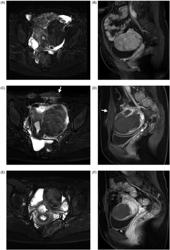
Table 2. The symptom changes at 6 months after treatment (n = 12).
Figure 3. A 24-year-old woman with a right broad ligament uterine fibroid. The CE-MRI showed a right broad ligament uterine fibroid oppressing the right iliac vessels (A) and the non-perfused area of ablated fibroid (97.8% of NPV ratio) with intact serosal layer after HIFU ablation (B).