Figures & data
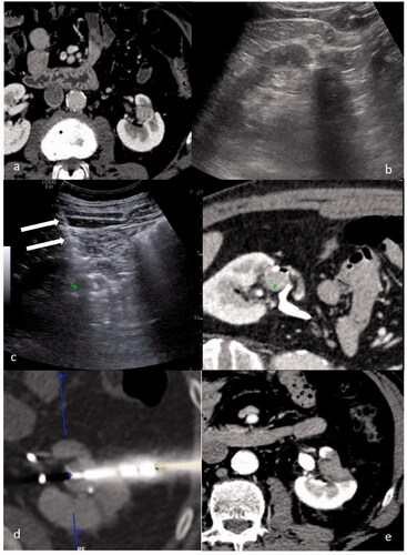
Figure 1. A case of a 52 y.o. patient with a single 12 mm RCC relapse after robotic assisted partial nephrectomy: (a) CT scan showing a 12 mm enhancing lesion in the left kidney. (b) US evaluation performed before treatment didn’t allow for clear identification of the tumor. (c) US/CT fusion imaging obtained during the treatment allowed to place a marker (green marker) on the tumor on the CT images to identify the target on US, and thus to provide guidance for needle insertion (white arrows). (d) CT scan performed immediately after needle insertion confirmed the correct targeting of the tumor (blue marker). (e) CT performed 24 h after the procedure showing a complete ablation.