
Figures & data
Figure 1. (a) Lobar Y90 administration in multifocal lesions. (b) Segmental Y90 administration in lesions confined to two or fewer hepatic segments.
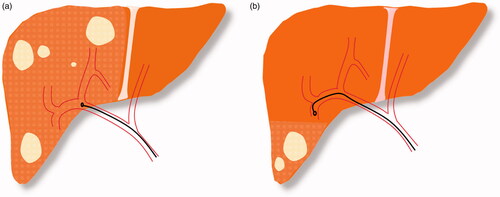
Figure 2. A 86 year-old male CRC patient with metastatic lesion in the left lobe (a) Pretreatment MRI showing baseline low-attenuation lesion in segment II/III measuring 6.3 × 3.2 cm (arrow). Radiation segmentectomy was performed for the patient and a dose of 235 Gy was administered. (b) MRI at one-year follow-up shows a shrunken non-enhancing tumor (3.6 × 1.9 cm, arrow) with retraction and atrophy of the treated segments (arrowhead).
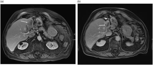
Table 1. Outcomes of trials evaluating the efficacy of radiation segmentectomy in colorectal liver metastasis.