Figures & data
Figure 1. Artificial right pleural effusion technique. The patient is placed in the left-lateral position, and pleural puncture is performed under aseptic technique through the right fourth intercostal space along the mid-axillary line using a 16-gauge epidural needle with Tuohy bevel (B., Braun Melsungen AG, Melsungen, Germany). Ventilation is suspended in the expiratory phase with an open adjustable pressure limit valve when the needle is introduced into the pleural cavity. Correct placement is confirmed when there is a passive loss of resistance to a falling column of fluid. Approximately 800 ml of warm normal saline is infused into the right pleural cavity.
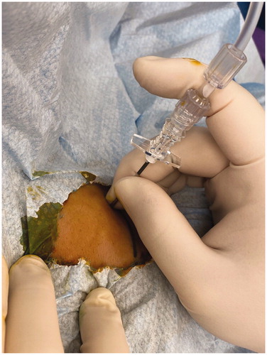
Figure 2. This patient with cirrhosis and HCC located in segment 7 of the liver being treated with HIFU. (A) CT image showing the location of the tumor (red arrow) which was in the liver dome and close to the diaphragm. (B) Tilting the HIFU transducer probe toward the cephalad direction, which is the right-hand side of this photo, in order to obtain an optimal view of the liver dome and the tumor. The patient is lying in the water bath in a right-lateral position. (C) HIFU sonographic image of the liver dome and the tumor (red arrow) obtained by this maneuver.
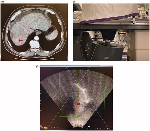
Figure 3. HIFU sonographic image of a patient with preexisting ascites and segment 6 HCC (red arrow). The patient is lying in the right-lateral position. The ascites has separated the liver dome (white arrow) from the diaphragm, allowing excellent sonographic visualization of the liver dome.
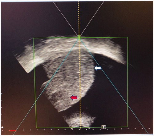
Figure 4. HIFU sonographic image of a small HCC in segment 8 (red arrow) being treated, after the instillation of 800 ml of normal saline into the right pleural cavity as artificial right pleural effusion (area denoted by ‘P’). A rib (denoted by ‘R’) is partially obscuring the liver in this intercostal view, therefore breath-holding is mandatory during sonication.
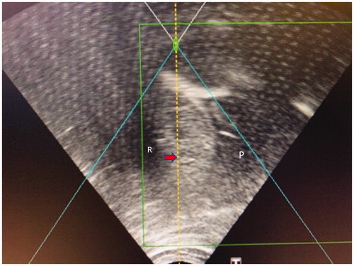
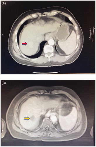
Figure 5. A 66-year-old man with cirrhosis and HCC in segment 7 of the liver. (A) Contrast CT scan showing the hypervascular tumor (red arrow). The patient subsequently underwent HIFU ablation of the tumor, facilitated by artificial right pleural effusion. (B) Contrast MRI taken 1 month after treatment shows no arterial enhancement in the tumor (yellow arrow), which is in keeping with good treatment response.