Figures & data
Table 1. Baseline characteristics of study patients.
Figure 1. A 62-year-old man with painful osteolytic T3 metastases from lung adenocarcinoma was treated with microwave ablation combined with bone cementing. Preoperative sagittal MRI (A), axial MRI (B), and axial CT (C) revealed osteolytic T3 metastases with the complete destruction of the posterior vertebral wall, tumor compression of the dural sac, and severe compression fracture. Using the left costal joint approach, a microwave antenna (arrow) was guided to the anterior edge of the lesion (D) by a bone puncture needle (arrow). After microwave ablation, 3 ml of bone cement was injected slowly (E), and the sagittal CT showed that the bone cement was deposited in the lesion area without obvious leakage (F). CT: computed tomography; MRI: magnetic resonance imaging.
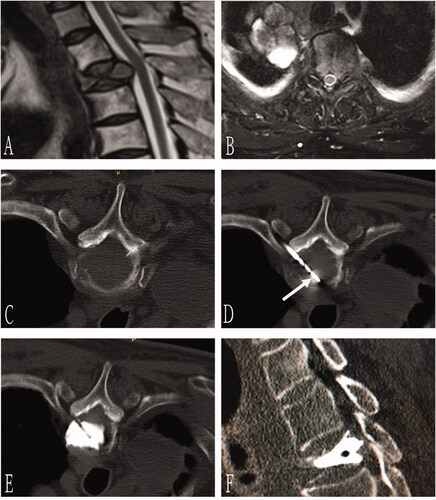
Figure 2. A 70-year-old man with T3 osteolytic metastases from lung adenocarcinoma was treated with microwave ablation combined with bone cement plasty. Preoperative axial CT showed T3 osteolytic destruction (A). The needle was inserted through bilateral costal vertebra joints, and the tip of the bone puncture needle was located in the middle 1/3 of the anterior vertebra (B). Next, the microwave antenna (arrow) was inserted through the right coaxial bone puncture needle (C), and 3 ml of bone cement was injected after microwave ablation (D). Sagittal (E) and coronal 3D CT (F) showed well-deposited bone cement in the vertebral body without extravasation.
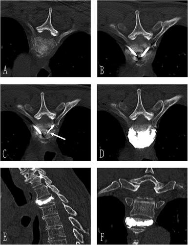
Table 2. Operative characteristics.
Figure 3. VAS score before and after the procedure. VAS: Visual Analog Scale. The VAS scores showed statistical significance pre- and post-MWA combined with PVP (p < 0.05).
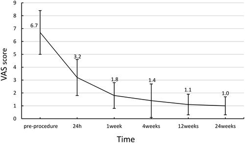
Figure 4. Daily morphine dose before and after the procedure. The daily morphine dose showed statistical significance pre- and post-MWA combined with PVP (p < 0.001).
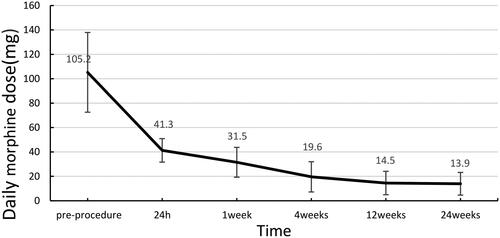
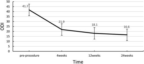
Figure 5. ODI score before and after the procedure. ODI: Oswestry Disability Index. The ODI scores showed statistical significance pre- and post-MWA combined with PVP (p < 0.05).