
Figures & data
Figure 1. Flow chart of patient enrollment.
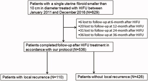
Figure 2. Measurement of distance from ventral side of fibroid to skin, distance from dorsal side of fibroid to skin, distance from dorsal surface of fibroid to sacrum, and the abdominal wall thickness.
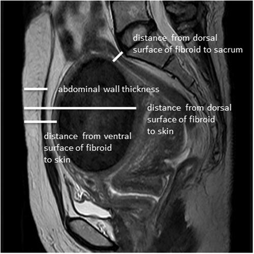
Figure 3. T2-weighted imaging signal intensity of uterine fibroids: (A) homogeneous hypointense. (B) homogeneous isointense. (c) homogeneous hyperintense. (D) heterogeneous hyperintense.
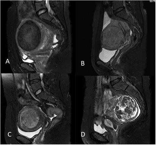
Figure 4. The degree of contrast enhancement of uterine fibroids: (A) mild enhancement. (B) moderate enhancement (myometrial grade). (C) significant enhancement.
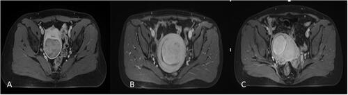
Figure 5. Contrast-enhanced MRI obtained from a patient with uterine fibroids in recurrent group. (A) One day post-USgHIFU image showed a NPV ratio of 62% was achieved. (B) 6-month post-USgHIFU image showed local recurrence of the treated fibroid.
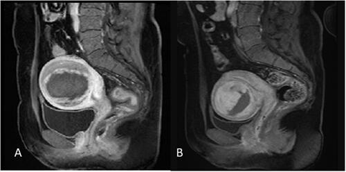
Figure 6. Contrast-enhanced MRI obtained from a patient with uterine fibroids in non-recurrence group. (A) One day post-USgHIFU imaging showed the fibroid was completely ablated. (B) 6-month post-USgHIFU imaging showed a shrinkage of 30% of the treated fibroid without local recurrence.
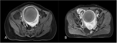
Table 1. Comparison of baseline characteristics of patients between the group with recurrence and group without recurrence.
Table 2. Comparison of HIFU treatment results between the group with recurrence and the group without recurrence.
Table 3. Binary logistic regression analysis results of independent factors affecting recurrence.