Figures & data
Figure 1. Structures of FeSO4-PyED and FeSO4-PyBD. FeSO4-PyED is capable of undergoing Bergman cyclization [Citation19], while FeSO4-PyBD is a cyclized ‘radical control’ version of FeSO4-PyED that does not undergo Bergman cyclization or form diradical intermediates.
![Figure 1. Structures of FeSO4-PyED and FeSO4-PyBD. FeSO4-PyED is capable of undergoing Bergman cyclization [Citation19], while FeSO4-PyBD is a cyclized ‘radical control’ version of FeSO4-PyED that does not undergo Bergman cyclization or form diradical intermediates.](/cms/asset/bca51b5e-0c10-46b6-aad2-db77ff416141/ihyt_a_2024280_f0001_b.jpg)
Figure 2. Effect of the diradical-generating enediyne moiety on enhancement of metalloenedyine cytotoxicity under hyperthermic conditions. U-1 cells were treated with various concentrations of FeSO4-PyED or its nonradical-generating analog, FeSO4-PyBD, for 1 h at 37 °C or 42.5 °C, and then plated for assessment of clonogenic survival and determination of surviving fraction. For FeSO4-PyBD data, error bars represent the standard error of the mean (SEM) from two experiments; FeSO4-PyED data are from one experiment that is representative of data from previously published experiments [Citation32].
![Figure 2. Effect of the diradical-generating enediyne moiety on enhancement of metalloenedyine cytotoxicity under hyperthermic conditions. U-1 cells were treated with various concentrations of FeSO4-PyED or its nonradical-generating analog, FeSO4-PyBD, for 1 h at 37 °C or 42.5 °C, and then plated for assessment of clonogenic survival and determination of surviving fraction. For FeSO4-PyBD data, error bars represent the standard error of the mean (SEM) from two experiments; FeSO4-PyED data are from one experiment that is representative of data from previously published experiments [Citation32].](/cms/asset/f205b255-d3ac-4c39-91ad-9f1a012700c6/ihyt_a_2024280_f0002_b.jpg)
Figure 3. FeSO4-PyED is a DNA-binding metalloenediyne. SYBR Green-labeled salmon sperm DNA was incubated in either water or various concentrations of FeSO4-PyED. Loss of dye fluorescence is reflective of binding (intercalation) of FeSO4-PyED to DNA. Error bars represent the SEM for triplicate determinations.
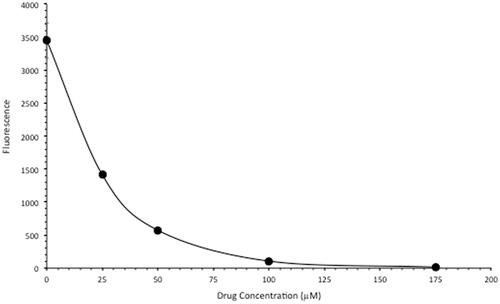
Figure 4. Differential effect of hyperthermia on apoptosis induced by FeSO4-PyED in U-1 melanoma and PIG1 melanocytes. Cells were incubated with 100 µM FeSO4-PyED or vehicle (1.5% water) for 1 h at 37 °C or 42.5 °C and then incubated at 37 °C with fresh media for various times. Apoptotic fractions were determined from bivariate plots of Annexin V-EGFP and PI fluorescence after treatments (not shown). The dashed line is representative of data from previously-published experiments with FeSO4-PyED on U-1 melanoma cells. Error bars represent SEM for 2–3 experiments.
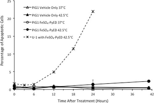
Figure 5. Treatment of C32 melanomas with FeSO4-PyED during heating inhibits tumor growth in a xenograft mouse model: Tumor volume measurements. Tumors were grown in the right hind limb of NRG mice, which were then injected with drug or vehicle and maintained for 1 h at either 33.5° or 42.5 °C by immersing the hind limb in a 34° or 43.5 °C water bath, respectively. Over the 14-day period of observation after treatment, tumor volume every other day was calculated and compared to the tumor volume on Day 0 (pre-treatment) to determine percent growth of each tumor. For each day that tumors were measured, the percent growth of each tumor was averaged with the other tumors in the same group to obtain a mean value of percent tumor growth. From days 5–14 post-treatment, One-way ANOVAs showed a significant difference between the groups (p = 0.001, days 7, 10, 12, and 14; p = 0.01 day 5; n = 10–13 mice). LSD post-hoc analyses showed significant differences (p < 0.05) between the FeSO4-PyED + Heat group and all other groups from days 12–14, and significant differences between the FeSO4-PyED + Heat group and the Vehicle + Heat and Vehicle Only groups on days 5–10 (p < 0.05) (or for p < 0.1, between the FeSO4-PyED + Heat group and all other groups from days 7 to 14). Data from three experiments (2–5 mice per group per experiment).
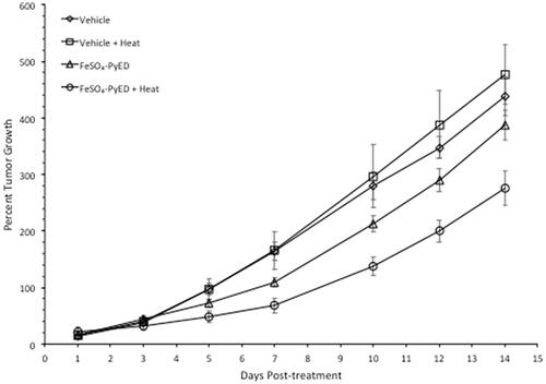
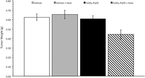
Figure 6. Treatment of C32 melanomas with FeSO4-PyED during heating inhibits tumor growth in a xenograft mouse model: Measurement of tumor weights. Tumor weights were determined on Day 14 after sacrificing mice that had been treated at either 33.5° or 42.5 °C with drug or vehicle as described in . Tumors obtained from mice treated with FeSO4-PyED during hyperthermia treatment were significantly smaller than the tumors in all other groups. The p value from a One-way ANOVA was p = 0.002. LSD post-hoc tests showed that tumor weights in the FeSO4-PyED + heat group was significantly different from tumor weights in each of the other groups (vs. Vehicle, p = 0.003; vs. Vehicle + Heat, p = 0.001; vs. FeSO4-PyED, p = 0.004; n = 10–13). Data from three experiments as noted in .