Figures & data
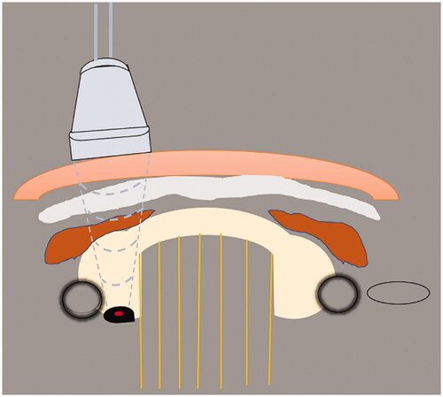
Figure 1. Schematic diagram of ultrasound-guided percutaneous thermal ablation of HPT. 1. Linear array probe used for guiding and positioning; 2. ablation needle; 3. skin and subcutaneous soft tissue; 4. fascia tissue; 5. anterior cervical muscles; 6. thyroid gland; 7. trachea; 8. carotid artery; 9. hyperplastic parathyroid gland; 10. internal jugular vein.
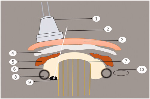
Table 1. Baseline characteristics of patients undergoing laser ablation for hyperparathyroidism.
Table 2. Baseline characteristics of patients undergoing radiofrequency ablation for hyperparathyroidism.
Figure 2. Sonogram of parathyroid adenoma before and after MWA. A 56-year-old woman was diagnosed with a parathyroid adenoma by ultrasound-guided biopsy before ablation. (a) Sonogram of parathyroid adenoma before MWA: the adenoma was hypoechoic, with a regular shape and clear boundary. Color Doppler flow imaging reveals a slightly rich-colored blood flow in the interior and edge; (b) Before MWA, the adenoma showed uniform and high enhancement in contrast-enhanced ultrasound (CEUS); (c) During ablation. (d) After ablation, there was almost no enhancement in CEUS. (e) One month after MWA, no obvious blood flow was found in the tumor.
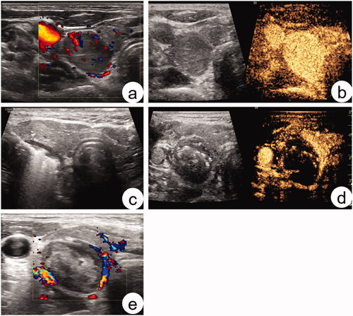
Figure 3. Schematic diagram of HIFU in the treatment of HPT.