
Figures & data
Figure 1. Sagittal MRI of fibroid at baseline (craniocaudal measurement 34.7 mm) and at 6 months post treatment (26.7 mm).
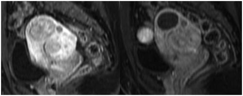
Figure 2. Axial MRI of fibroid at baseline (36.1 × 47.4 mm) and at 6 months post treatment (33 × 35.8 mm).
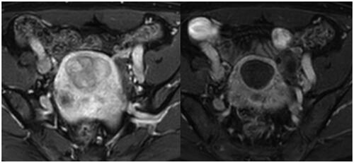
Figure 3. Consort Flow Chart.
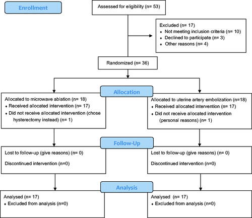
Figure 4. Line plot of volume change (ml) before and after treatment. MWA: Microwave ablation; UAE: Uterine artery embolization.
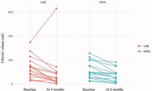
Table 1. Baseline characteristics.
Table 2. Primary and Secondary outcomes.
Supplemental Material
Download PDF (271.4 KB)Data availability statement
Will individual participant data be available (including data dictionaries)? Yes- upon reasonable request.
What data in particular will be shared? All available data in the database will be made available.
What other documents will be available? No other information will be made available.
When will data be available (start and end dates)? Data is saved for 10 years and is then destroyed.
By what access criteria will data be shared. (including with whom, for what types of analyses, and by what mechanism)? Data will be made available upon reasonable request as judged by the research group.