
Figures & data
Table 1. Demographic data of patients in both groups.
Figure 1. (a) A 63-year-old male CRLM patient with the primary tumor origin in the colon. The patient did not have extrahepatic metastases and was assigned to the patient group with curative clinical indication. T2-weighted (a) and T1-weighted contrast-enhanced (b) MRI images before MWA show three hepatic metastases in segments 4a, and 7. CT images (c, d and e) show the position of the Antenna during the ablation procedure. Follow-up T1-weighted contrast-enhanced MRI at six months (f) show complete ablation and no recurrence at the site of ablation.
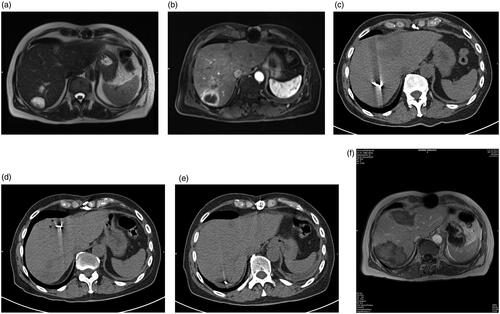
Table 2. Overview of survival according to the volume, diameter and energy output.
Figure 2. Median survival rate for curative and palliative indication. This figure describes the median survival rate for 57 CRLM patients with curative and 75 CRLM patients with palliative treatment indications. The Kaplan–Meier estimator was created using the BiAS program. The survival rate is shown in percent on the y-axis. The x-axis describes the survival times in days.
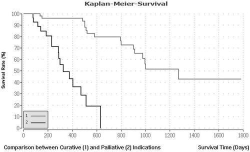
Figure 3. Median survival rate for all patients. Kaplan–Meier curve of the median monitoring rate for 132 CRLM patients with curative and palliative treatment indications. All microwave ablations were performed between 2010 and 2017.
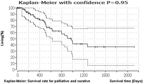
Table 3. Overview of survival according to the primary tumor origin.