
Figures & data
Figure 1. Flowchart of population enrollment.
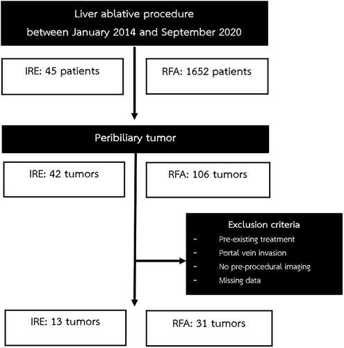
Table 1. Demographic data and tumor characteristics.
Figure 2. Pre-operative, intra-operative, and post-operative imaging of IRE group. (A) Fifty-one-year-old man with a small hepatocellular carcinoma at liver segment IV. (A) The arterial phase MRI of liver showed a 25-mm arterial enhancing nodule less than 5 mm from the bile duct. (B) The axial non-contrast enhanced CT during the IRE procedure revealed the proper position of the two IRE electrodes. (C) The immediate post-operative contrast-enhanced CT showing the ablated area of the tumor. (D) A 1-month follow-up MRI revealed complete IRE ablation without residual tumor but with adjacent focal bile duct dilatation (arrow).
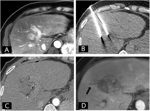
Figure 3. Local tumor progression-free survival in the IRE and RFA groups.
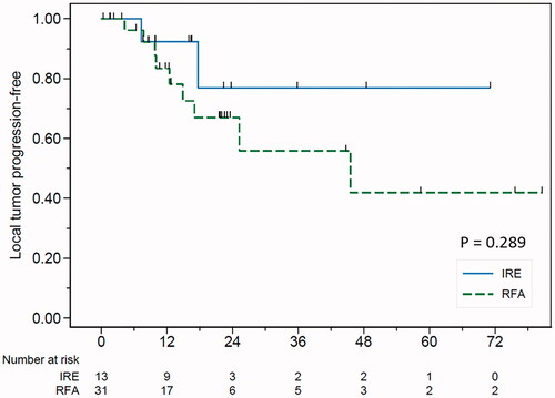
Figure 4. Biliary complication with clinically significant liver atrophy after the RFA procedure. (A) Seventy-six-year-old woman with a small hepatocellular carcinoma. (A) Contrast-enhanced CT of the liver in the portal phase showed an 11-mm washout nodule abutting the bile duct (white arrow). (B) Realtime sonography was performed to confirm the position of the RFA electrode. (C) A 1-month follow-up contrast-enhanced CT of the liver shows complete ablation with segmental bile duct dilatation (arrow). (D) A 5-month follow-up revealed lobar atrophy of the whole left lobe of liver (arrowhead) representing a major bile duct and vascular pedicle injury.
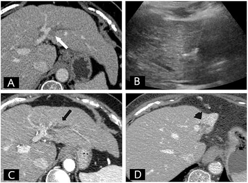
Table 2. Outcomes and complications of the procedure.
Table 3. Multivariate analysis of predictive factors for biliary complications.