
Figures & data
Figure 1. Flowchart of the study population.
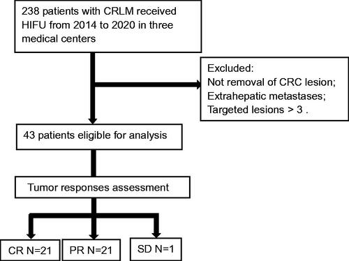
Figure 2. An example of partial response for HIFU ablation. The patient is a 54-year-old male with liver metastasis from rectal cancer. The contrast-enhanced MRI showed that pre-procedure hepatic lesions at segment VIII and segment I invaded the vena cava and hepatic vein (a) and the non-perfused area occupied more than 70% of the whole tumor (white arrow) after HIFU ablation (b).
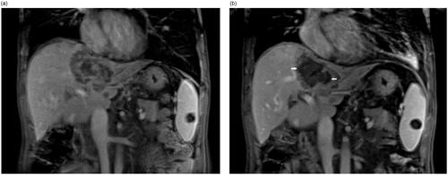
Figure 3. An example of a complete response for HIFU ablation. The patient is a 54-year-old female with liver metastasis from colon cancer. The contrast-enhanced MRI showed that pre-procedure hepatic lesion located between 1st and 2nd porta (white arrow) (a) and complete non-perfused area (white arrow head) of the whole tumor after HIFU ablation (b).
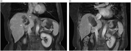
Figure 4. Kaplan–Meier survival curves for comparison between the CR and PR of patients with CRLM after USgHIFU treatment, and median survival was 35 months and 23 months in the CR patients and PR patients, respectively (p = 0.00).
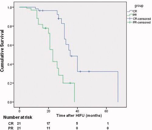
Table 1. Characteristics of patients with CRLM treated by USgHIFUa.