
Figures & data
Figure 1. ATO at low concentrations does not inhibit the proliferation of HCC cells after insufficient RFA. HCC cells were treated with ATO and MTT assay was performed to assess the inhibitory effects on cell proliferation. (A) SMMC7721or HepG2 was treated with ATO for 72 h and IC50 was calculated. (B) SMMC7721 and SMMC7721-H cells were treated with ATO at 1, 2, and 3.5 μM concentrations. The differences in inhibitory effects of ATO on SMMC7721 and SMMC7721-H cells are shown. (C) HepG2 and HepG2-H cells were treated with ATO at 1, 2, and 3.5 μM concentrations. The differences in inhibitory effects of ATO on HepG2 and HepG2-H cells are shown. At least three independent experiments were performed. ns, no significance; *p<0.05; **p<0.01; and ***p<0.001.
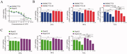
Figure 2. HCC cells after insufficient RFA promote tube formation of endothelial cells by paracrine manner, and ATO suppresses the process. CM was collected from HCC cells or HCC cells after insufficient RFA with or without ATO treatment and used to treat endothelial cells. CM from (A) SMMC7721 or SMMC7721-H cells and (B) HepG2 or HepG2-H cells, with or without ATO treatment was used to treat endothelial cells and tube formation was examined. (C) Statistical results of (A) and (B) are shown. At least three independent experiments were performed. ns, no significance; *p<0.05; **p<0.01; and ***p<0.001.
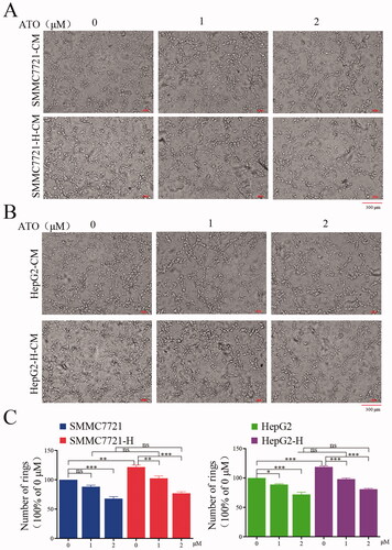
Figure 3. ATO inhibits angiogenesis induced by HCC cells after insufficient RFA through the Ang-1/Ang-2/Tie2 pathway. (A, B) The concentration of Ang-1 and Ang-2 in conditioned media from SMMC7721 and SMMC7721-H, or HepG2 and HepG2-H cells, with or without ATO treatment was measured. (C) The expression of Ang-1 or Ang-2 in HCC cells and HCC cells after insufficient RFA was examined using Western blot. (D, E) Ang-1 or Ang-2 was knocked down in SMMC7721-H or HepG2-H cells, CM was collected, and tube formation was assessed. (F) CM from untreated or RFA-treated HCC cells with or without ATO treatment was used to treat endothelial cells, and p-Tie2 expression was analyzed. (G) Ang-1 or Ang-2 was knocked down, CM was used to treat endothelial cells, and p-Tie2 expression was analyzed. (H, I) Tie2 was knocked down in endothelial cells, and the effect of CM from HCC cells on tube formation was examined. At least three independent experiments were performed. *p<0.05; **p<0.01; ***p<0.001; and ns: no significance.
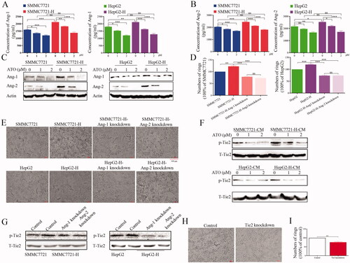
Figure 4. Hif-1α overexpression abolishes the inhibitory effect of ATO on angiogenesis after insufficient RFA. (A) HCC cells were treated with ATO and the expression of Hif-1α and p-Akt was analyzed. (B) HCC cells were treated with YC-1 and the expression of Hif-1α, p-Akt, Ang-1, and Ang-2 was analyzed. (C) The concentrations of Ang-1 and Ang-2 in CM after YC-1 treatment were measured in HCC cells after insufficient RFA. (D) HCC cells were treated with YC-1 and p-Tie2 expression was analyzed. (E) Western blot was used to validate the transfection efficacy of plasmid containing Hif-1α. (F) After Hif-1α overexpression, HCC cells were treated with ATO, CM was collected and used to treat endothelial cells, and p-Tie2 expression was analyzed. (G) HCC cells with Hif-1α overexpression were treated with ATO and the expression of p-Akt, Hif-1α, Ang-1, and Ang-2 was analyzed. (H, I) HCC cells with Hif-1α overexpression were treated with ATO and the concentrations of Ang-1 and Ang-2 in CM were determined. At least three independent experiments were performed. BC: Blank control; NC: Negative control; and Over: Overexpression. *p<0.05; **p<0.01; ***p<0.001; and ns: no significance.
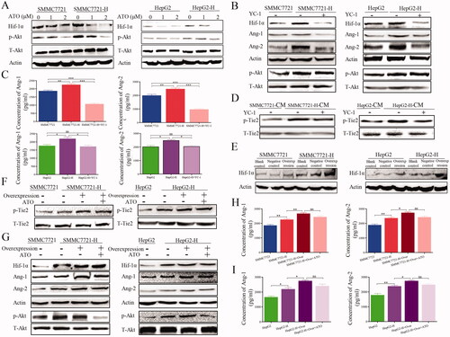
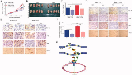
Figure 5. ATO restrains tumor growth and angiogenesis in HCC after insufficient RFA. (A) SMMC7721 and SMMC7721-H cells were subcutaneously inoculated into nude mice, which were then treated with PBS or ATO. After four weeks, the animals were sacrificed. Tumor size was measured with a caliper after every other day. (B) Images representing tumor volume at the end of experiments, n = 5 per group. (C) Tumor weight was measured at the end of the experiment. (D) Tumor sections were stained for the detection of Hif-1α, Ang-1, Ang-2, or CD31. Representative images of immunohistochemical analysis. (E) Representative images with high magnification. (F) Quantification of CD31. (G) Schematic shows the effect of ATO on endothelial cells and the underlying mechanisms, in HCC after insufficient RFA. At least three independent experiments were performed. *p<0.05; **p<0.01; and ns: no significance.
Data availability statement
The datasets used and analyzed during the study are available from the corresponding author upon reasonable request.