Figures & data
Table 1. Patient characteristics and treatment details.
Table 2. Outcomes based on nodule volume change and clinical results.
Figure 1. Changes in iPTH, serum calcium levels, nodule volume, and volume reduction ratio (VRR) of the three groups before and after hyperplastic parathyroid gland radiofrequency ablation (RFA) at each follow-up. The iPTH in PHPT and SHPT both showed successful treatment responses compared with baseline (A) even though both SHPT subgroups showed a transient rebound (B). The calcium level in PHPT and SHPT both decreased at follow-up duration (C), but SHPT groups experienced transient hypocalcemia, especially in the SHPT without PTX subgroup (D). Nodule volumes in all three groups significantly decreased over time after RFA (E). The VRR in PHPT and SHPT without PTX were both over 95% and significantly higher than SHPT with PTX (F). *p < 0.05 versus baseline.
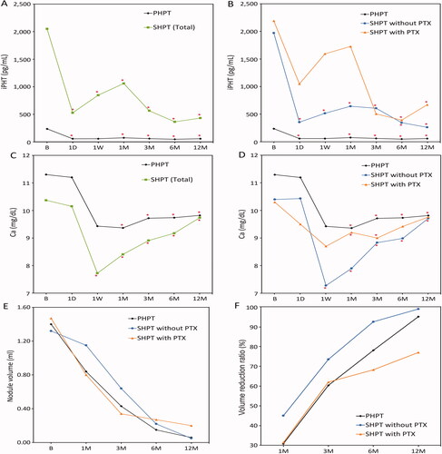
Table 3. Laboratory outcomes.
Figure 2. A 54-year-old female with primary hyperparathyroidism. Technetium (99mTC)-sestamibi scan (A) and US examination (B) revealed a hyperfunctional left lower parathyroid adenoma measuring 14.5 mm × 6.3 mm. CT scan of the parathyroid adenoma (arrow) without (C) and with contrast enhancement before RFA (D). CT scan without (E) and with contrast enhancement 1 year after RFA (F) showed total regression (arrow) of the left inferior parathyroid adenoma. Serum PTH and calcium levels were decreased and remained in the normal range at the 1-year follow-up. RFA: radiofrequency ablation; PTH: parathyroid hormone; US: ultrasound.
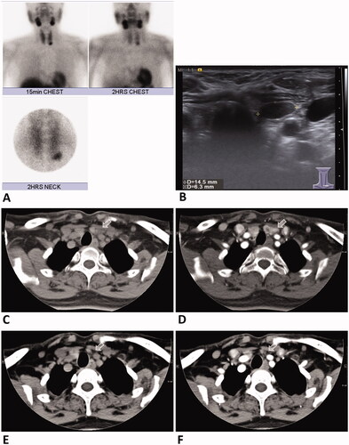
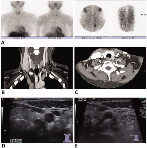
Figure 3. A 41-year-old female with secondary hyperparathyroidism status post parathyroidectomy. Technetium (99mTC)-sestamibi scan (A) revealed a left inferior hyperparathyroid nodule, with high signal also shown on forearm due to parathyroid tissue autotransplantation postparathyroidectomy. Contrast-enhanced CT scan of coronal (B) and axial view (C) revealed a hyperplastic parathyroid nodule in the left inferior neck (arrow). US examination (D) revealed a left inferior parathyroid hyperplastic nodule measuring 8.6 mm × 7.3 mm. US examination follow-up at 4 months after RFA (E) showed significant decrease in size of the nodule measuring 6.1 mm × 5.2 mm. Serum PTH and calcium levels were decreased at 3-month follow-up after treatment. RFA: radiofrequency ablation; PTH: parathyroid hormone; US: ultrasound.