Figures & data
Figure 1. Axial MRI scan showing diffuse T2-weighted hyperintense tissue in the left pleural cavity (white intrapleural rim), extending to the left fissure and pericardium, being highly suspicious for pleural extension of previous peritoneal PMP.
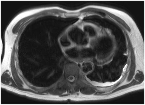
Figure 2. The pleura is transformed into a fibrous tissue containing irregular mucinous pools. These may contain slightly atypical cilindrical epithelium (white arrow), either free-floating in the mucin (A) or still attached to the fibrous wall of the pool (B).
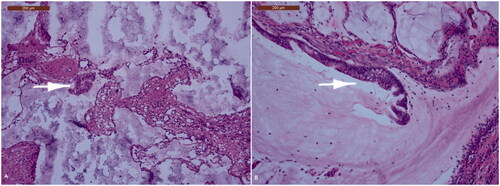
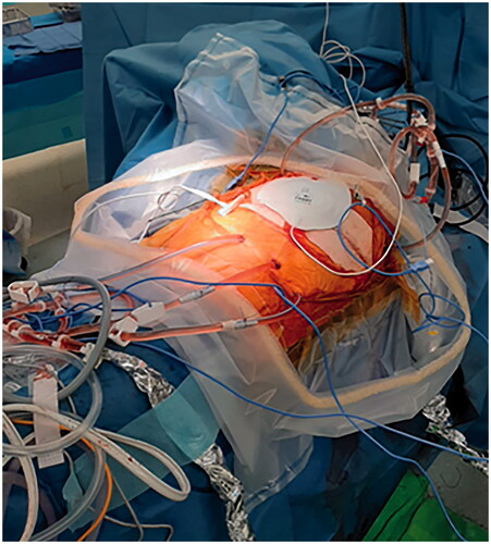
Figure 3. Intra-operative image of HITHOC using 2 inflow (Ch24) and 3 outflow cannulas (Ch28). The left pleural cavity was perfused for 30 min at 41 °C using 4700 cc of Dianeal and oxaliplatin (460 mg/m2).