
Figures & data
Table 1. Comparison of baseline characteristics of CSP patients in three types.
Figure 1. The signal of blood flow and contrast-enhanced ultrasound obtained from a patient with type III CSP treated with HIFU. A/C: abundant blood supply around the embedding area of the GS in the previous cesarean section scar (white arrow) before HIFU ablation; B/D: reduced blood supply around the embedding area of the GS in the previous cesarean section scar (white arrow) after HIFU ablation.
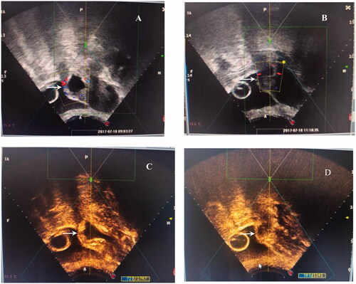
Figure 2. A. Transabdominal ultrasound image (sagittal view) of Foley catheter balloon (black arrow) inflated with 20 ml fluid and used for hemostasis by compression at the time of CSP treatment. B. The end of Foley catheter was connected with a drainage bag to calculate the amount of bleeding.
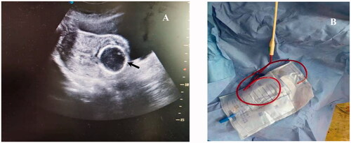
Table 2. Comparison of HIFU treatment results for CSP patients in three types.
Table 3. Comparison of Ultrasound-guided suction curettage results and follow-up in three groups.
Table 4. Correlation between the type of CSP and parameters of HIFU treatment and ultrasound-guided-suction curettage (n = 153).