
Figures & data
Figure 1. A representative ultrasound image of cervical pregnancy. The red arrow is showing a gestational sac, and the white arrow is showing off the germ. The gestation is located in the cervical canal, but the uterine cavity was empty (yellow arrow).
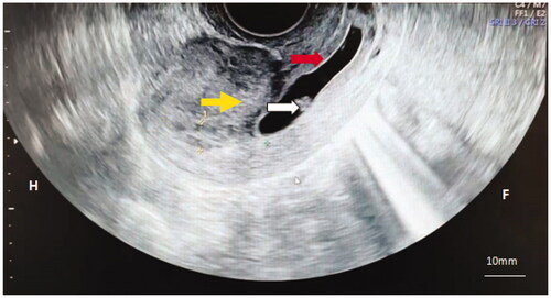
Figure 2. Color flow Doppler imaging demonstrated successful removal of the gestational tissue. (A) Contrast-enhanced Ultrasound was performed before HIFU, which shows of obvious perfusion of the gestational sac existed (white arrow). (B) Contrast-enhanced Ultrasound was performed again after HIFU treatment, and it shows no obvious perfusion agent was detected in the gestational sac tissue (white arrow).
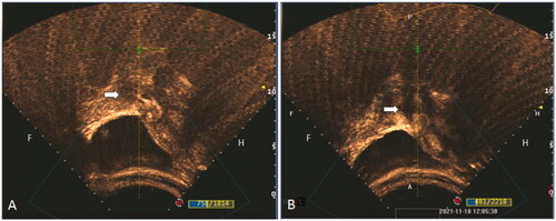
Table 1. Baseline clinical characteristics of seven patients with cervical pregnancy treated with HIFU combined with HGSC.
Table 2. Ultrasound parameters of HIFU treatment on seven patients.
Table 3. Clinical parameters of suction curettage for seven patients.
Table 4. Follow-up observations after HIFU and HGSC treatments.