Figures & data
Figure 1. B-Ultrasound for patient before HIFU (Aug. 17th, 2020). The B-Ultrasound for patient showed the mass is 61.20 × 18.70 × 48.10 mm.
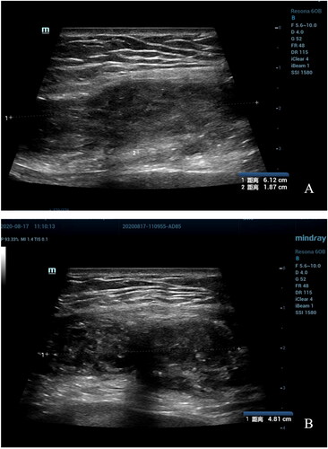
Figure 2. MRI for patient before HIFU (Aug. 22nd, 2020). (A) Sagittal T2-weighted image showed the mass’s size is 63.7 × 21.0 mm; (B) Post-contrast sagittal T1-weighted image showed the mass’s size is 64.6 × 25.2 mm; (C) Axial T2-weighted image showed the mass’s size is 63.9 mm.
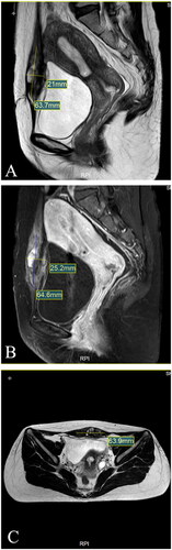
Figure 3. HIFU Treatment (Sept. 04th, 2020). The image showed the mass during HIFU.
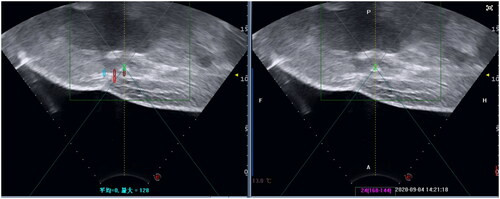
Figure 4. MRI for patient in 1st day after HIFU (Sept. 04th, 2020). (A) Sagittal T2-weighted image showed the mass’s size is 79.0 × 22.4 mm; (B) Post-contrast sagittal T1-weighted imae showed the mass’s size is 70.4 × 20.1 mm; (C) Axial T2-weighted image showed the mass’s size is 70mm.
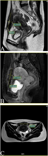
Figure 5. MRI for patient in 3rd month after HIFU (Dec. 11st, 2020). (A) Sagittal T2-weighted image showed the mass’s size is 10.6 × 5.8 mm; (B) Post-contrast sagittal T1-weighted image showed the mass’s size is 10.9 × 5.5 mmI; (C) Axial T2-weighted image showed the mass’s size is 8.3 mm.
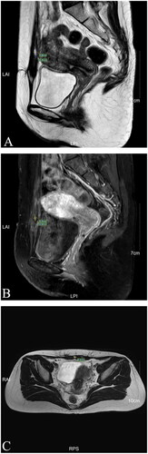
Figure 6. MRI for patient in 7th month after HIFU (Apr. 20th, 2021). (A) Sagittal T2-weighted image showed the mass’s size is 4.4 mm; (B) Post-contrast sagittal T1-weighted image showed the mass’s size is 5mm; (C) Axial T2-weighted image showed the mass’s size is 4.8 mm.
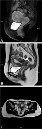
Figure 7. MRI for patient in 1st year after HIFU (Dec. 17th, 2021). (A) Sagittal T1-weighted image showed the mass’s size is 4.1 mm; (B) Post-contrast sagittal T1-weighted image showed the mass’s size is 3.9 mm; (C) Axial T2-weighted image showed the mass’s size is 4.8 mm.
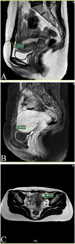
Figure 8. B-Ultrasound for patient in 2nd year after HIFU (Jun. 12nd, 2022). The B-Ultrasound showed no abnormalities about two years after HIFU.
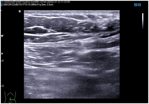
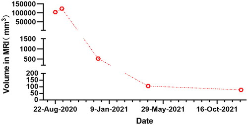
Figure 9. Shrinkage of the AWE mass volume after HIFU treatment.