
Figures & data
Figure 1. MMWA for a patient with adenomyosis who had anemia. (a) Diagram of trans-abdominal ultrasound-guided ablation of the myometrial lesion. The ‘mobile-layered ablation’ technique was adopted to move the ablation needle from deep to superficial positions in a fan-shaped ablation zone; (b) preoperative contrast-enhanced ultrasonography showed hyper-enhancing lesions in the arterial phase; (c) ultrasound-guided ablation of myometrial lesions with a thermal field of hyperechoic signals around the ablation needle (white arrow); (d) postoperative contrast-enhanced ultrasonography reveals no perfusion in the myometrial ablation area.
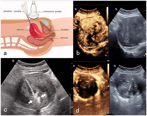
Figure 2. MEWA for an adenomyosis patient with anemia. (a) Diagram of trans-abdominal ultrasound-guided the myometrial and endometrial ablation. An ablation needle was inserted into the endometrium near the uterine fundus, and the ablation area was one-third of the endometrium near the uterine fundus. (b) Transabdominal ultrasound-guided insertion of the ablation needle into the endometrium near the uterine fundus. (c) Hyperechoic signals are detected immediately after the endometrium ablation (white arrow). (d) Post-ablation imaging reveals no perfusion of the upper one-third of the endometrium near the uterine fundus (white arrow).
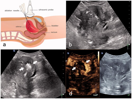
Table 1. Comparison of preoperative clinical baseline characteristics between the two groups.
Table 2. Clinical efficacy regarding various monitoring indicators pre- and postoperatively in the MMWA group.
Table 3. Clinical efficacy regarding various monitoring indicators pre- and postoperatively in the MEWA group.
Figure 3. (a–d) Changes in Hb, CA125, uterine volume, uterine-volume reduction in MMWA and MEWA groups.
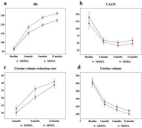
Figure 4. (a–d) Changes in SSS, HRQL, VAS, and MFS in MMWA and MEWA groups.
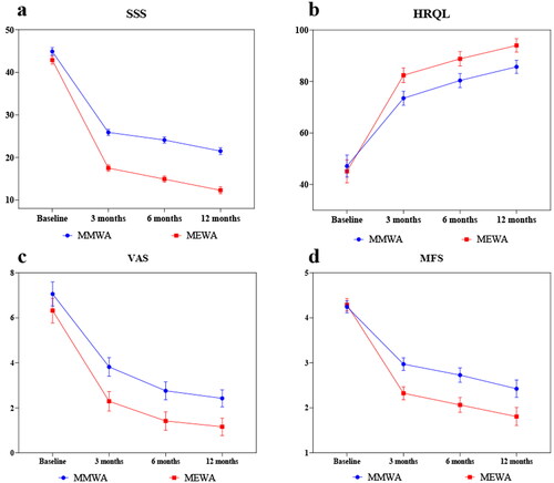
Table 4. Comparison of clinical efficacy between the two groups.