
Figures & data
Figure 1. Ablation of uterine myomas using different ultrasound-guided techniques. A. Transabdominal ultrasound guidance. B. Combined transabdominal and transvaginal ultrasound guidance.
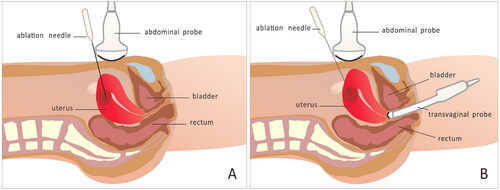
Table 1. Comparison of clinical baseline data before treatment for uterine myomas in the TA group and the TA/TV group.
Figure 2. TA/TV US-guided PMWA for the treatment of UM. A. TAU disturbed by strong echo, in which the needle tip is not clearly displayed (white arrow). B. TVU is not disturbed by strong echo, with clear display of the needle tip (white arrow). C. The strong echo generated by ablation under TAU monitoring interferes with the display of the posterior tissue. D. Clear display of the posterior tissue and part of uterine serosa in TVU monitoring. TA/TV: combined transabdominal and transvaginal; US: ultrasound; PMWA: percutaneous microwave ablation; UM: uterine myoma; TAU: transabdominal ultrasound; TVU: transvaginal ultrasound
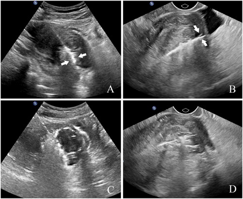
Figure 3. US-guided establishment of artificial ascites and PMWA treatment for UM. A–B. Artificial ascites (white stars) form a water barrier between the uterus and surrounding tissue structures, allowing a clear display of the uterine serosa. US: ultrasound; PMWA: percutaneous microwave ablation; UM: uterine myoma.
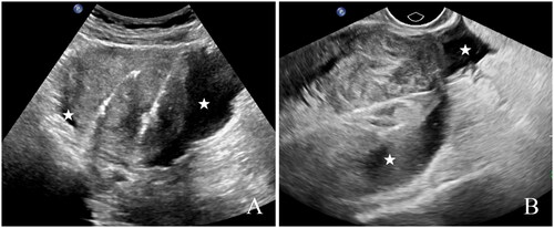
Table 2. Image display of the same lesion in the same patient in the TA/TV group according to guidance technique.
Table 3. Intraoperative supplementary ablation, postoperative efficacy and safety comparison of PMWA treatment for uterine myomas according to different guidance technique.